Marta Brigid Maggioni1, Martin Krämer1,2, and Jürgen R. Reichenbach1,2,3,4,5
1Medical Physics Group, Institute of Diagnostic and Interventional Radiology, Jena University Hospital - Friedrich Schiller University, Jena, Germany, 2Institute of Diagnostic and Interventional Radiology, Jena University Hospital - Friedrich Schiller University, Jena, Germany, 3Michael Stifel Center for Data-driven and Simulation Science Jena, Friedrich Schiller University, Jena, Germany, 4Abbe School of Photonics, Friedrich Schiller University, Jena, Germany, 5Center of Medical Optics and Photonics, Friedrich Schiller University, Jena, Germany
1Medical Physics Group, Institute of Diagnostic and Interventional Radiology, Jena University Hospital - Friedrich Schiller University, Jena, Germany, 2Institute of Diagnostic and Interventional Radiology, Jena University Hospital - Friedrich Schiller University, Jena, Germany, 3Michael Stifel Center for Data-driven and Simulation Science Jena, Friedrich Schiller University, Jena, Germany, 4Abbe School of Photonics, Friedrich Schiller University, Jena, Germany, 5Center of Medical Optics and Photonics, Friedrich Schiller University, Jena, Germany
The proposed undersampled AFI method reduces the acquisition time of a AFI-based B1 map from 30 to 2 min in vivo, while still providing robust and consistent VFA-based AFI-corrected T1 estimations.
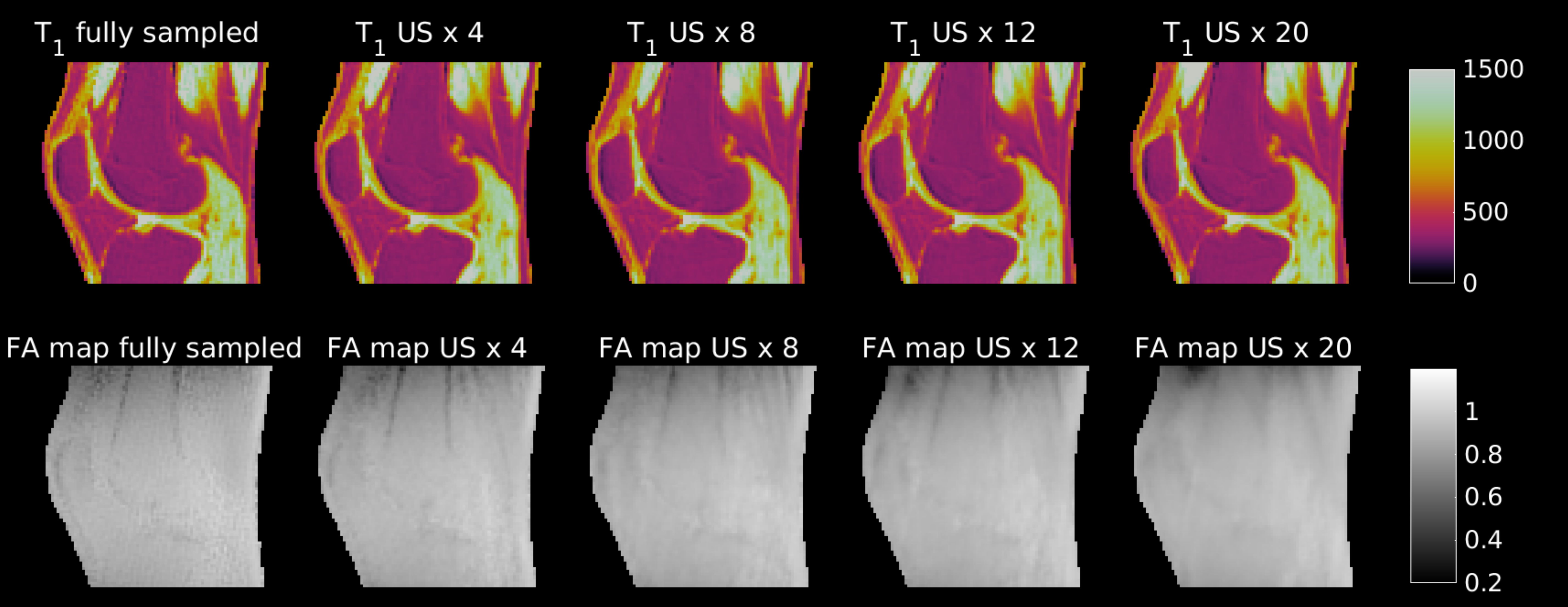
Figure 2: The top row presents T1 maps (in ms) of the knee of a volunteer after B1 correction with increasingly undersampled AFI-based B1 maps (bottom row).
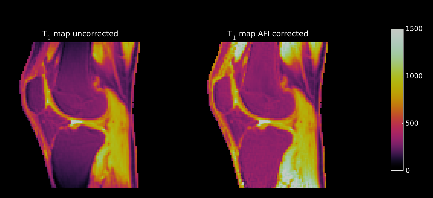
Figure 1: T1 maps of the knee of a volunteer before and after the B1 correction with fully sampled AFI-based B1 maps. Notice the drop of the signal at the edge of the FoV that is corrected by the B1 map. The images are scaled between 0 and 1500 ms.