Céline Smekens1, Quinten Beirinckx2, Floris Vanhevel3, Pieter Van Dyck3, Arjan den Dekker2, Jan Sijbers2, Thomas Janssens1, and Ben Jeurissen2
1Siemens Healthcare NV/SA, Beersel, Belgium, 2imec-Vision Lab, Department of Physics, University of Antwerp, Wilrijk, Belgium, 3Department of Radiology, Antwerp University Hospital and University of Antwerp, Edegem, Belgium
1Siemens Healthcare NV/SA, Beersel, Belgium, 2imec-Vision Lab, Department of Physics, University of Antwerp, Wilrijk, Belgium, 3Department of Radiology, Antwerp University Hospital and University of Antwerp, Edegem, Belgium
Super-resolution T2* mapping based on UTE Spiral VIBE MRI allows for high-resolution T2* mapping of knee structures, showing comparable T2* maps to maps based on direct 3D UTE Spiral VIBE acquisitions while requiring approximately 25% less scan time.
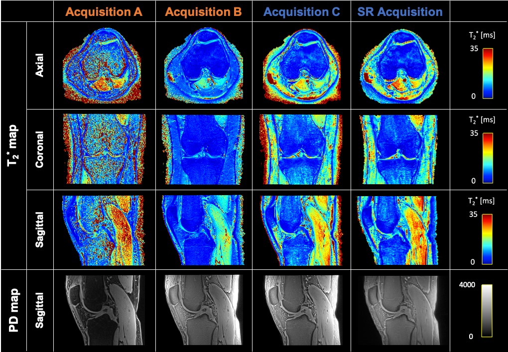
Figure 3 – Representative T2* and PD maps estimated from 2 short (A and B) and 2 long (C and SR) acquisitions.
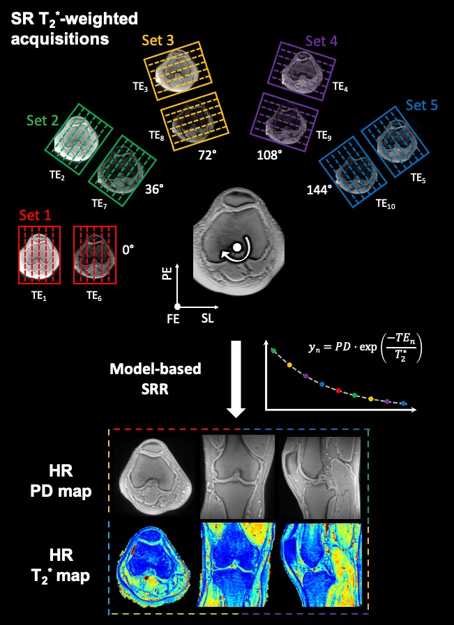
Figure 1 – Schematic representation of the super-resolution (SR) T2*-weighted acquisitions and model-based super-resolution reconstruction (SRR). 5 UTE Spiral VIBE datasets, consisting of 2 TEs each, were acquired with high in-plane and low through-plane resolution, while rotating around the frequency-encoding axis over angles of 0°, 36°, 72°, 108° and 144°. A model-based SRR framework, including a mono-exponential T2* relaxation model, was used to estimate high-resolution (HR) proton density (PD) and T2* maps directly from the series of low resolution T2*-weighted images.