Marco Fiorito1, Maksym Yushchenko1, Davide Cicolari2, Mathieu Sarracanie1, and Najat Salameh1
1Center for Adapatable MRI Technology (AMT center), Department of Biomedical Engineering, University of Basel, Allschwil, Switzerland, 2Department of Physics, University of Pavia, Pavia, Italy
1Center for Adapatable MRI Technology (AMT center), Department of Biomedical Engineering, University of Basel, Allschwil, Switzerland, 2Department of Physics, University of Pavia, Pavia, Italy
Diagnosis
and treatment monitoring can benefit from local $$$T_1$$$ information. Here, a fast
Look-Locker based $$$T_1$$$ mapping sequence is used to produce an in vivo map
of a volunteer’s hand at 0.1 T.
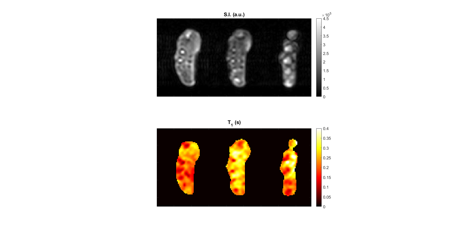
In vivo $$$T_1$$$ map of a
volunteer’s hand. The main structures visible in the reference anatomical
image (2D GRE) are retrieved in the $$$T_1$$$ map. In this case, a shorter TI (25 ms)
was chosen to better characterise the structures with a short $$$T_1$$$ .
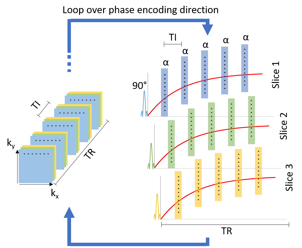
Schematic representation of
the Look-Locker-based $$$T_1$$$ mapping sequence. The interleaved scheme allows to
change the number of slices without impacting the scan time. Nonetheless, more
slices signify longer TIs, which can impact the retrieval of short relaxation
times. The use of a saturation pulse was
chosen to avoid waiting for full $$$T_1$$$ recovery, hence reducing the acquisition
time.