Kaia Ingerdatter Sørland1, Pål Erik Goa2,3, Kirsten Margrete Selnæs1,3, Elise Sandsmark3, Cristopher George Trimble1, Mohammed R. S. Sunoqrot1, Mattijs Elschot1,3, and Tone F. Bathen1,3
1Department of Circulation and Medical Imaging, Norwegian University of Science and Technology, Trondheim, Norway, 2Department of Physics, Norwegian University of Science and Technology, Trondheim, Norway, 3Department of Radiology and Nuclear Medicine, St. Olavs University Hospital, Trondheim, Norway
1Department of Circulation and Medical Imaging, Norwegian University of Science and Technology, Trondheim, Norway, 2Department of Physics, Norwegian University of Science and Technology, Trondheim, Norway, 3Department of Radiology and Nuclear Medicine, St. Olavs University Hospital, Trondheim, Norway
Pseudo-T2 values achieved with Autoref normalization of prostate T2-weighted images are comparable to the gold standard prostate T2 values obtained with T2 mapping. The T2 value contrast between the prostate zones can also be conserved with Autoref normalization.

Table 1: The prostate T2 values from the multi-echo spin echo (MESE) imaging sequence and the prostate pseudo-T2 values from Autoref with different reference tissues, averaged over seven healthy volunteers.
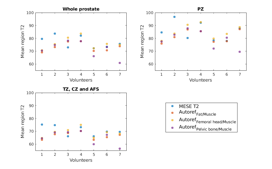
Figure 2: The spread in mean prostate T2 and pseudo-T2 for all seven volunteers, both from MESE and Autoref with three pairs of reference tissues.