Yitong Li1, Xiaoqing Liang1, Bowen Hou1, Yan Xiong1, Weiyin Vivian Liu2, and Xiaoming Li1
1Tongji Hospital, Tongji Medical College, Huazhong University of Science and Technology, Wuhan, China, 2MR Research, GE Healthcare, Beijing, China
1Tongji Hospital, Tongji Medical College, Huazhong University of Science and Technology, Wuhan, China, 2MR Research, GE Healthcare, Beijing, China
Coils may influence quantitative measurements of
synthetic lumbar spine MRI; therefore, usage of the same coil should be adopted
when one study is carried out.

Figure 2: Box
plots for T1, T2, and PD values of different
tissues compared between different coils. VIS means
considering the vertebral bodies and intervertebral discs together. * indicates
p <0.05; ** indicates p <0.01; *** indicates p <0.001.
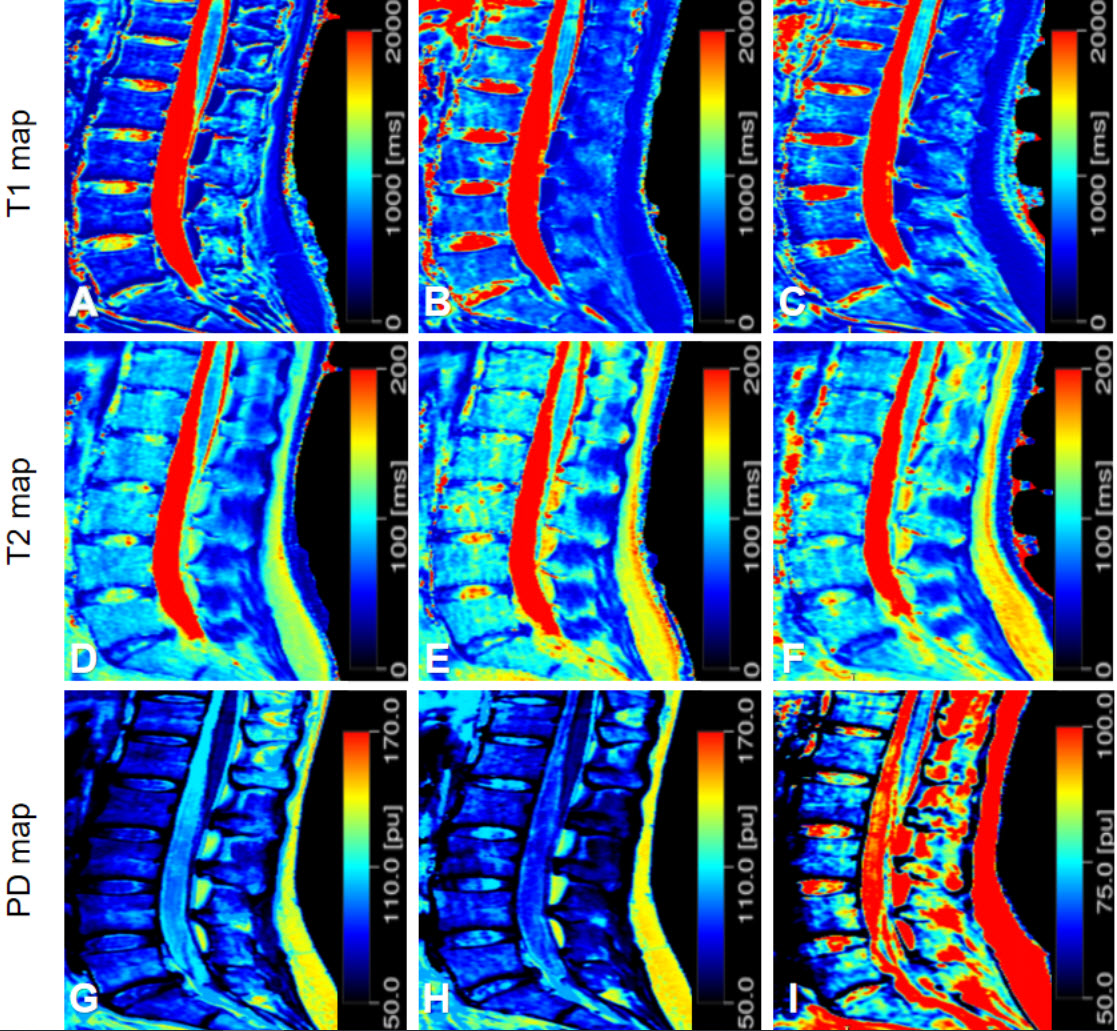
Figure
1: Quantitative maps of a 26-year-old female volunteer. (A-C) T1 map; (D-F) T2
map; (G-I) PD map. Maps from left to right were obtained using Spine DST, Body,
and Flex Large, respectively.