Ping Wang1, Jay D Turner1, Juan Uribe1, and John C Gore2
1Barrow Neurological Institute, Phoenix, AZ, United States, 2Institute of Imaging Science, Vanderbilt University, Nashville, TN, United States
1Barrow Neurological Institute, Phoenix, AZ, United States, 2Institute of Imaging Science, Vanderbilt University, Nashville, TN, United States
A T1ρ dispersion imaging method has been successfully developed for human lumbar spine. This technique has potential for detecting proteoglycan loss in the early degenerative disc disease.
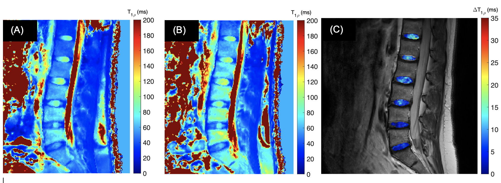
Fig. 3: T1ρ maps acquired under FSL = 100Hz (A) and 300Hz (B), with TSLs = [1ms, 11ms, 21ms, 31ms, 41ms]. The T1ρ dispersion, i.e., T1ρ = T1ρ(300Hz) - T1ρ(100Hz), is displayed in (C).
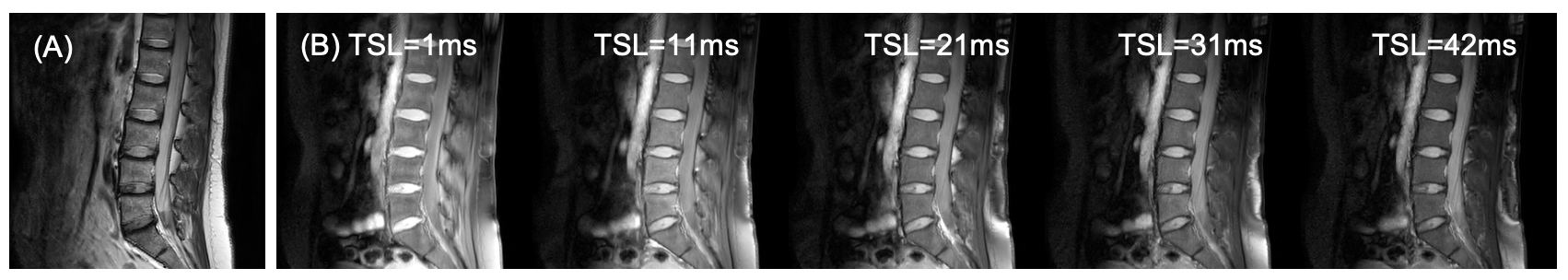
Fig. 2: T1ρ imaging on a 43-yrs female healthy volunteer. (A) T2-weighted image for structural information. (B) T1ρ-weighted images at spin-lock times (TSLs) = [1ms, 11ms, 21ms, 31ms, 41ms] under a spin-lock frequency (FSL) of 300Hz.