Kavita Singh1, Simone Cauzzo1,2, Maria Guadalupe Garcia Gomar1, Matthew Stauder1, Nicola Vanello3, Claudio Passino2,4, and Marta Bianciardi1
1Brainstem Imaging Laboratory, Department of Radiology, Athinoula A. Martinos Center for Biomedical Imaging, Boston, MA, United States, 2Sant'Anna School of Advanced Studies, Institute of Life Sciences, Pisa, Italy, 3Dipartimento di Ingegneria dell’Informazione, University of Pisa, Pisa, Italy, 4Fondazione Toscana Gabriele Monasterio, Pisa, Italy
1Brainstem Imaging Laboratory, Department of Radiology, Athinoula A. Martinos Center for Biomedical Imaging, Boston, MA, United States, 2Sant'Anna School of Advanced Studies, Institute of Life Sciences, Pisa, Italy, 3Dipartimento di Ingegneria dell’Informazione, University of Pisa, Pisa, Italy, 4Fondazione Toscana Gabriele Monasterio, Pisa, Italy
Using high spatial resolution 7 Tesla resting-state fMRI and a recently developed in-vivo brainstem nuclei atlas, we report the functional connectome of arousal and motor brainstem nuclei in living humans.
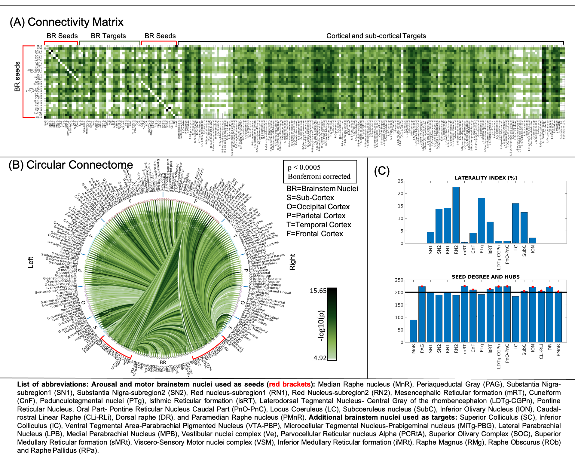
Figure1: (A) Connectivity matrix (i.e. mean connectivity values, n = 20) and (B) functional connectome (both thresholded at p < 0.0005, Bonferroni corrected for display purposes, 31 seeds and 195 targets) of arousal and motor brainstem nuclei (red brackets), exhibiting specific connectivity within the brainstem and with cortical and sub-cortical regions. (C) These nuclei showed high symmetry and high connectivity degree. Interestingly, 11 brainstem nuclei were network hubs: PAG, mRT, CnF, isRT, LDTg-CGPn, PnO-PnC, SubC, ION, CLi-RLi, DR and PMnR (see figure for abbreviations).
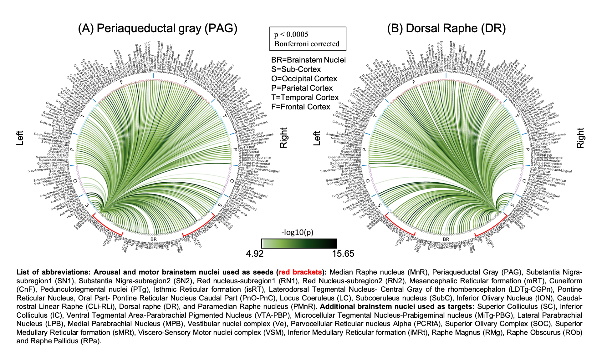
Figure 2. Functional connectome of the (A) Periaqueductal gray (PAG) and (B) Dorsal Raphe (DR) nucleus (p < 0.0005, Bonferroni corrected for display purposes, n =20). Literature8 review shows PAG connectivity to pre-frontal cortex, thalamus, insular cortex, cingulate cortex as seen in our results. LC, RPa, CnF, CLi-RLi, SC and IC also showed connectivity with PAG as expected, except for RMg, ROb, amygdala. DR showed connectivity to SN2, LC, LPB, MPB, VTA-PBP, PAG, caudate, putamen, thalamus, hippocampus and amygdala as reported in earlier studies8 (see figure for abbreviations).