André Döring1, Maryam Afzali1, Elena Kleban1, Roland Kreis2, and Derek K Jones1
1Cardiff University Brain Research Imaging Centre (CUBRIC), Cardiff University, Cardiff, United Kingdom, 2Departments of Radiology and Biomedical Research, University of Bern, Bern, Switzerland
1Cardiff University Brain Research Imaging Centre (CUBRIC), Cardiff University, Cardiff, United Kingdom, 2Departments of Radiology and Biomedical Research, University of Bern, Bern, Switzerland
An efficient Monte-Carlo simulation was
implemented for studying intracellular metabolite diffusion in realistic tissue
samples of glial cells. The effect of glial activation on non-Gaussian and anomalous diffusion was modeled to study the
resolution limit of diffusion MR spectroscopy.
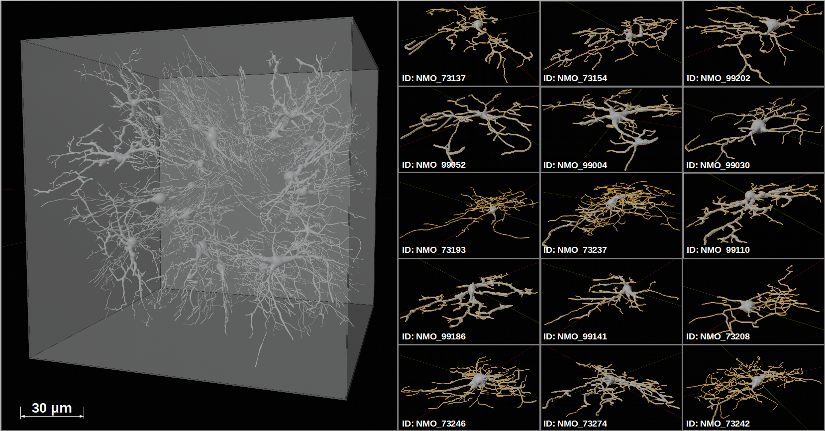
Fig. 2: Left:
The reference tissue structure (RTS) consists of 30 monkey glial cells in a VOI
of 150x150x150μm³. To create an isotropic tissue environment 15
different cells were placed twice, each randomly rotated, in the VOI. The
Blender physics engine was used to avoid cell collision and overlap. Right:
Overview of the 15 different glial cells [NeuroMorpho.org IDs are provided].
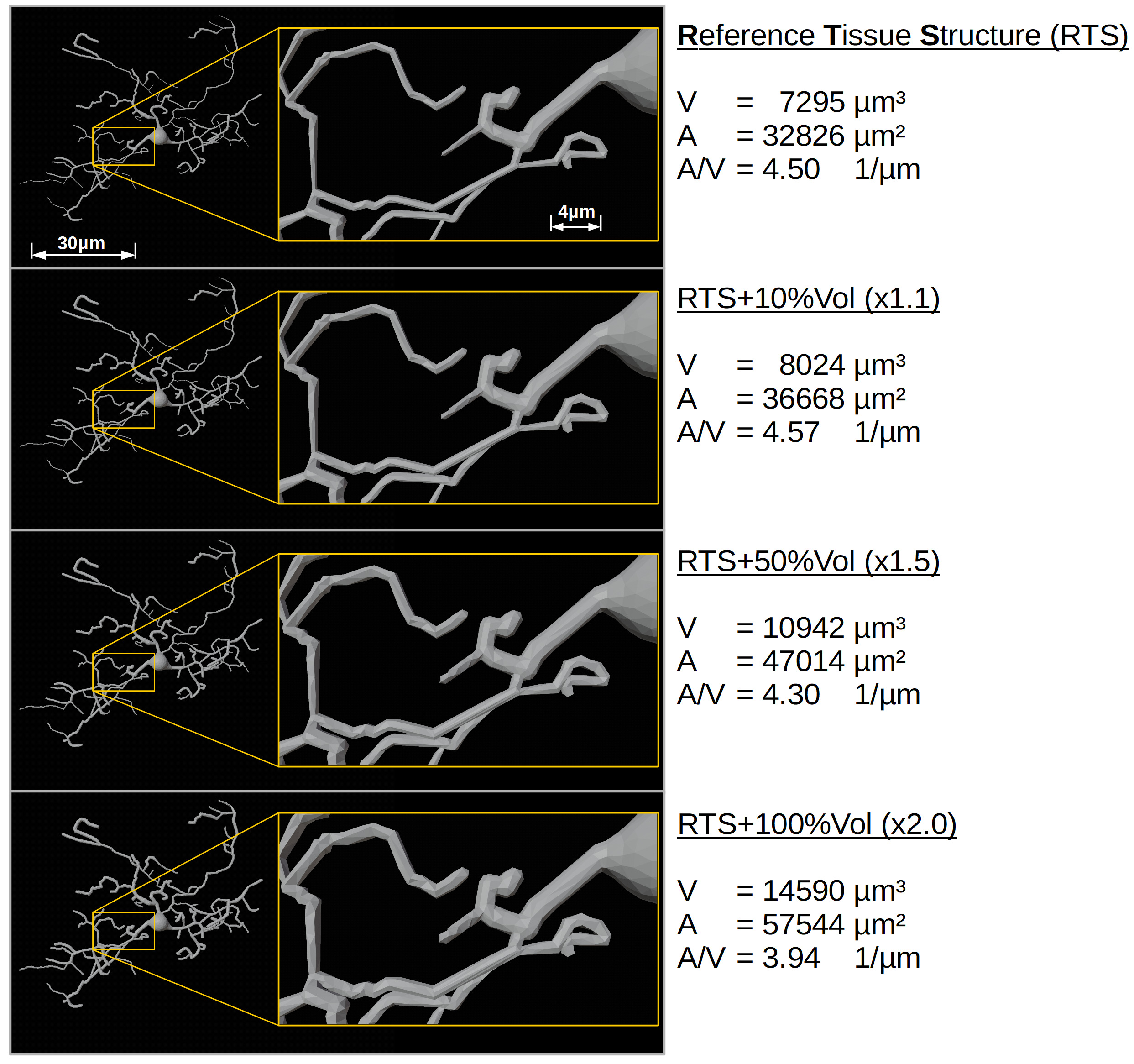
Fig. 3: Left: swelling of a glial cell (ID: 73137) with a
volume increase by 10%, 50% or 100% (zoomed insets to highlight differences).
Right: property changes of the RTS in volume (V), surface area (A) and surface area
to volume ratio (A/V) upon cell-swelling for the entire VOI presented in
Fig. 2.