Alicia Cronin1,2, Sarah Detombe3, Camille Duggal2, Neil Duggal3, and Robert Bartha1,2
1Medical Biophysics, University of Western Ontario, London, ON, Canada, 2Centre for Functional and Metabolic Mapping, Robarts Research Institute, London, ON, Canada, 3Clinical Neurological Sciences, University Hospital, London Health Sciences Centre, London, ON, Canada
1Medical Biophysics, University of Western Ontario, London, ON, Canada, 2Centre for Functional and Metabolic Mapping, Robarts Research Institute, London, ON, Canada, 3Clinical Neurological Sciences, University Hospital, London Health Sciences Centre, London, ON, Canada
This study found that degenerative
cervical myelopathy (DCM) patients with severe spinal cord compression also
demonstrated larger regions of cortical activation in the primary motor cortex
during a controlled finger-tapping task.
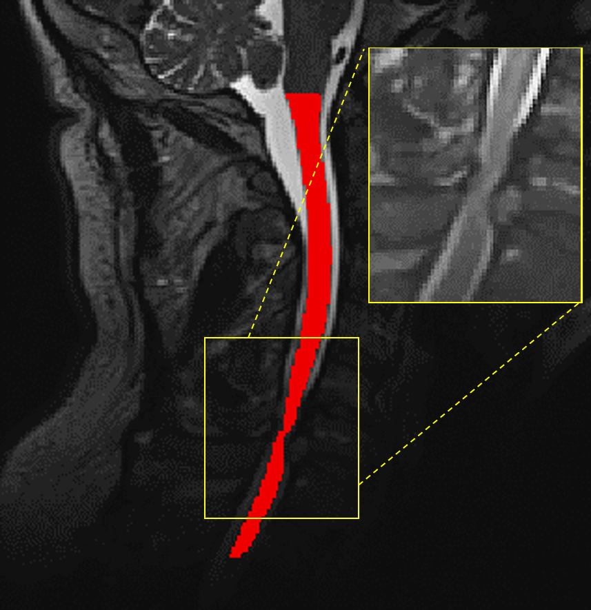
Figure 1: T2-weighted image of the cervical spinal cord of a DCM
patient showing the segmented cord in red and the compression site displayed on
the inset.
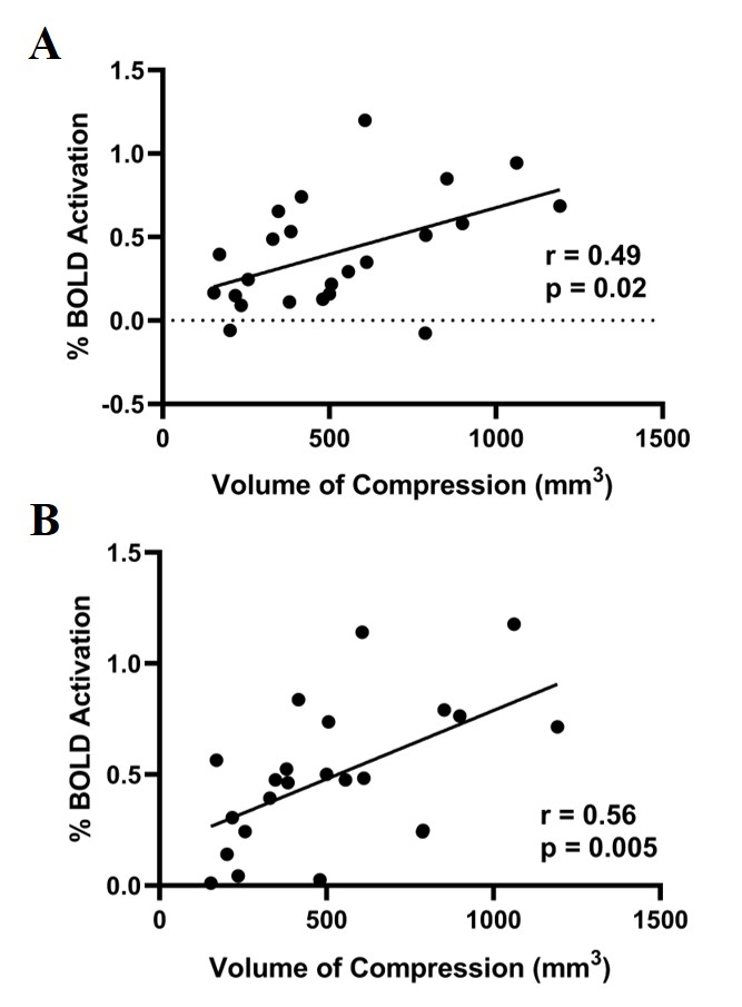
Figure 2: A: % BOLD signal correlation with spinal cord compression volume when tapping with left hand. B: % BOLD signal correlation with spinal cord compression volume when tapping with right hand.