Simon Lévy1,2,3, Patrick Freund4,5,6, Virginie Callot1,2,3, and Maryam Seif4,5
1CRMBM, Aix-Marseille University, CNRS, Marseille, France, 2CEMEREM, APHM, Hopital Universitaire Timone, Marseille, France, 3iLab-Spine International Research Laboratory, Marseille-Montreal, France, 4Spinal Cord Injury Center, University Hospital Balgrist, University of Zurich, Zurich, Switzerland, 5Max Planck Institute for Human Cognitive and Brain Sciences, Leipzig, Germany, 6Wellcome Trust Centre for Neuroimaging, UCL Institute of Neurology, London, United Kingdom
1CRMBM, Aix-Marseille University, CNRS, Marseille, France, 2CEMEREM, APHM, Hopital Universitaire Timone, Marseille, France, 3iLab-Spine International Research Laboratory, Marseille-Montreal, France, 4Spinal Cord Injury Center, University Hospital Balgrist, University of Zurich, Zurich, Switzerland, 5Max Planck Institute for Human Cognitive and Brain Sciences, Leipzig, Germany, 6Wellcome Trust Centre for Neuroimaging, UCL Institute of Neurology, London, United Kingdom
The Intra-Voxel Incoherent Motion technique at 3T has the ability to map spinal cord perfusion and depict gray matter, even on a single-subject basis, with sensitivity to capillary network orientations and fair inter-slice reproducibility. More patients data are needed and will be acquired.

Fig. 2: Mean IVIM parameters maps across HCs (N=11) at mid C1, C2, C3 levels in the template space (0.5mm isotropic, average of 15 slices) for each diffusion-encoding direction (A-P: anteroposterior, R-L: right-left, I-S: inferosuperior) and averaged across directions. The mean MEDIC image across subjects helps visualize GM and evaluate the quality of the template registration. Below, the mean signal profile is shown for each direction. The linear fit on high b-values helps visualize the deviation from the diffusion-only signal decay, illustrating the challenge along the I-S axis.
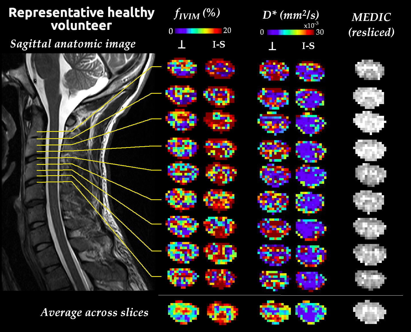
Fig. 3: Slice-wise fIVIM and D* maps in a representative healthy volunteer (30 years old man) for diffusion encoding in the transverse plane (⊥, mean across maps with A-P and R-L diffusion encoding) and along the I-S axis. The mean maps across slices (after registration based on the MEDIC middle slice image) are shown at the bottom. The slices position is displayed on the sagittal anatomic image (T2-weighted turbo spin-echo, 0.3×0.3mm2 in-plane resolution, 2.75mm slice thickness). The MEDIC image resliced to the IVIM data resolution helps visualize the GM shape along with the IVIM maps.
