Ying Chu1 and Jürgen Finsterbusch1
1Department of Systems Neuroscience, University Medical Center Hamburg-Eppendorf, Hamburg, Germany
1Department of Systems Neuroscience, University Medical Center Hamburg-Eppendorf, Hamburg, Germany
Partial-SMS
acceleration of combined fMRI acquisitions of the brain and cervical spinal
cord is feasible with a loss in image quality or SNR but can reduce the
acquisition time considerably.
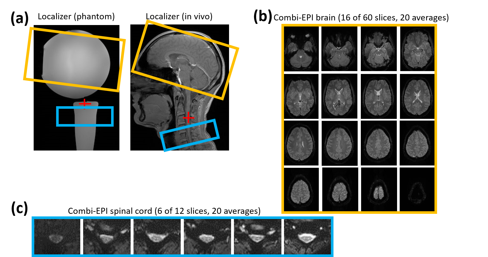
Figure 1: (a) Localizers
showing the typical geometric setup for cortico-spinal fMRI: the brain (orange)
and spinal cord (blue) volumes and the isocenter (red cross). (b, c) in vivo EPI
example images of the (b) brain (16 of 60 slices), and (c) spinal cord (6 of 12
slices) using SMS acceleration with a factor of 3 for the brain volume only.
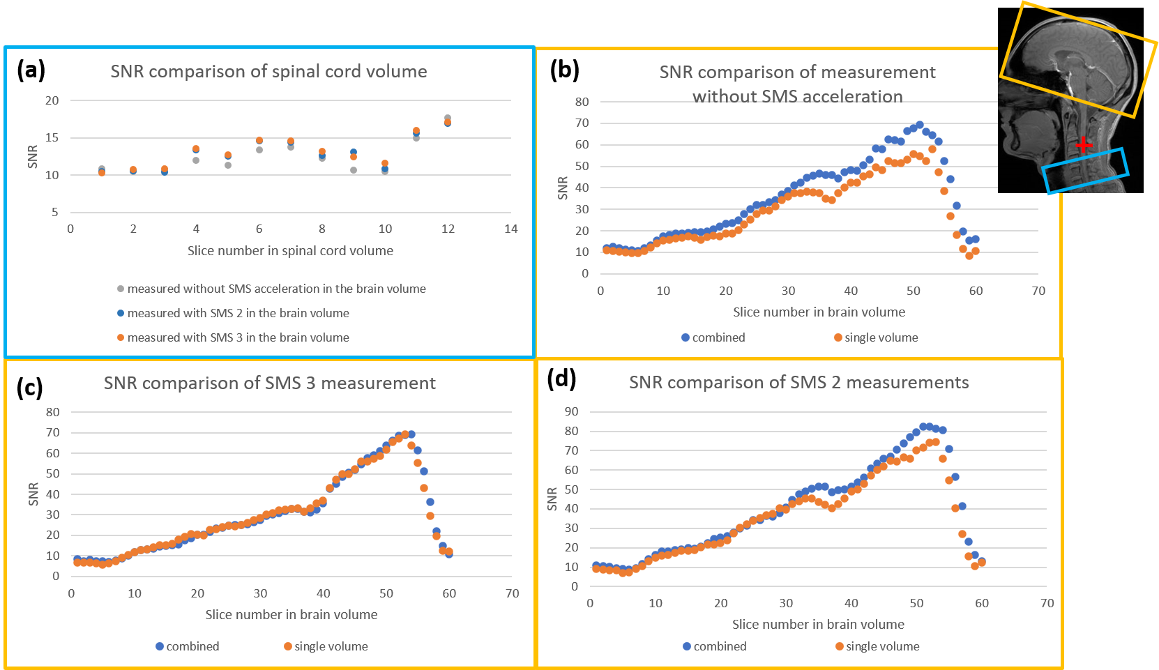
Figure 3: Results of SNR measurements on an in
vivo volunteer covering a brain volume only and combined brain and spinal cord volumes in the same acquisition: (a) spinal
cord without simultaneous multi-slice imaging (SMS) and (b-d) brain volume (b)
without SMS acceleration and (c,d) with SMS acceleration factors of 2 and 3,
respectively.