Junjie Ma1, Craig R. Malloy1,2,3, Crystal E. Harrison1, James Ratnakar1, Galen D. Reed4, Vlad G. Zaha1,2, and Jae Mo Park1,3,5
1Advanced Imaging Research Center, UT SOUTHWESTERN MEDICAL CENTER, Dallas, TX, United States, 2Internal Medicine, UT Southwestern Medical Center, Dallas, TX, United States, 3Radiology, UT Southwestern Medical Center, Dallas, TX, United States, 4GE Healthcare, Dallas, TX, United States, 5Electrical and Computer Engineering, UT Dallas, Richardson, TX, United States
1Advanced Imaging Research Center, UT SOUTHWESTERN MEDICAL CENTER, Dallas, TX, United States, 2Internal Medicine, UT Southwestern Medical Center, Dallas, TX, United States, 3Radiology, UT Southwestern Medical Center, Dallas, TX, United States, 4GE Healthcare, Dallas, TX, United States, 5Electrical and Computer Engineering, UT Dallas, Richardson, TX, United States
The
proposed method achieved the dual-phase acquisition for hyperpolarized 13C-labeled
metabolites for human heart. Two different
phases (end systole and end diastole) could be clearly distinguished for all HP
metabolites in short-axis and long-axis ventricle views.
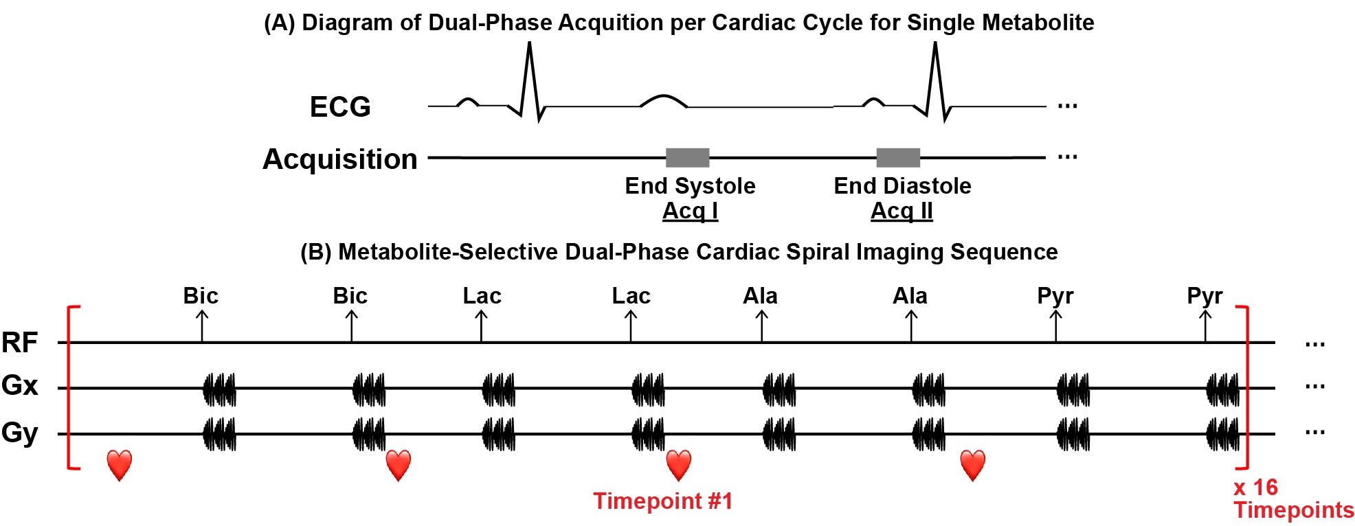
Figure 1. Acquisition scheme for each metabolite
and the proposed imaging sequence. (A) For
each metabolite, two acquisitions were conducted during each cardiac cycle,
which are at end systole and end diastole, respectively. (B) Multi-echo
images for hyperpolarized [13C]bicarbonate, [13C]lactate,
[1-13C]alanine and [1-13C]pyruvate are acquired in order
with the interval of 1 R-R for each timepoint. Images from in total 16
timepoints are acquired.
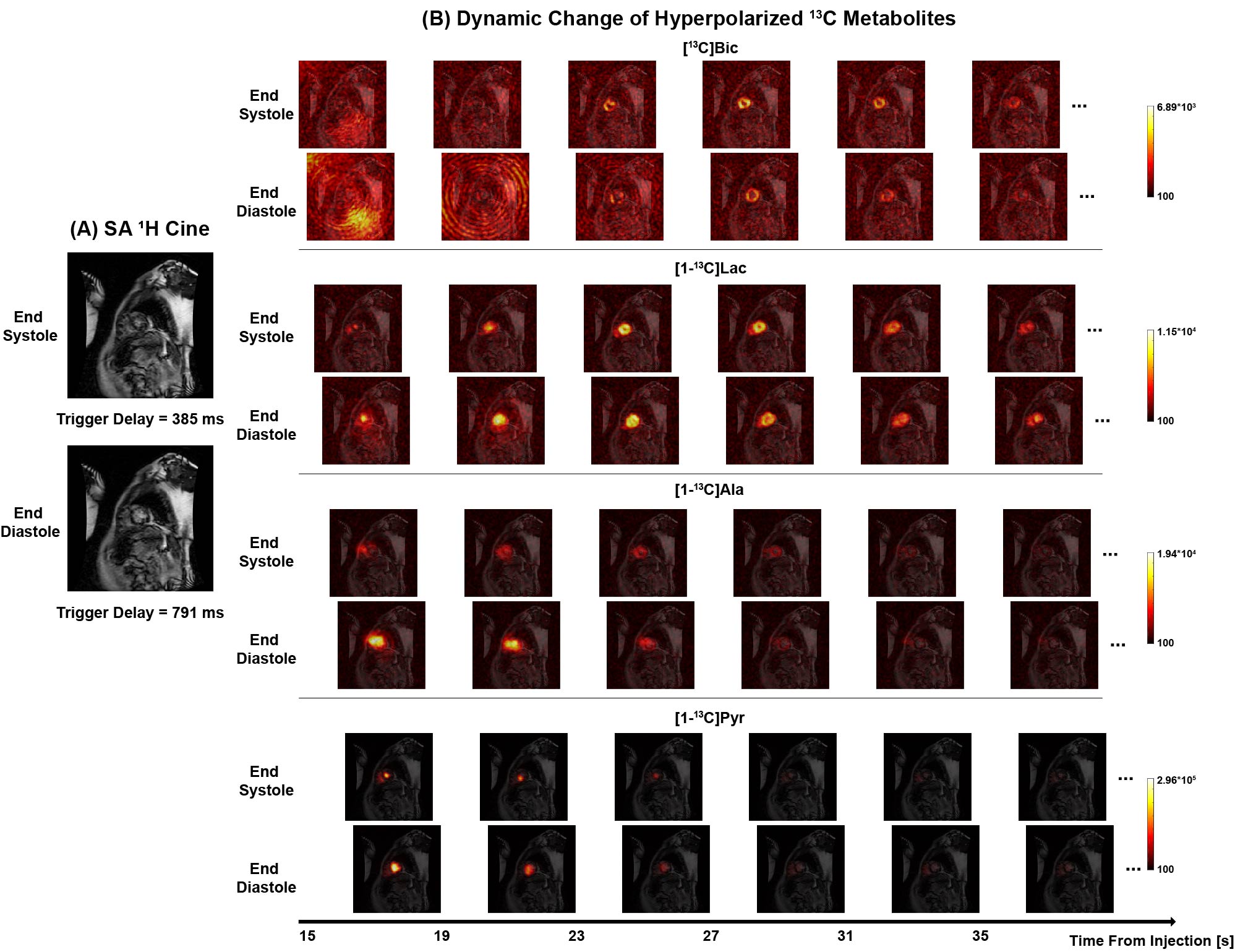
Figure 3. Dual-phase hyperpolarized [1-13C]pyruvate
cardiac imaging in SA plane. (A) 1H cardiac images in SA plane with the trigger delays of 385 ms (end systole)
and 791 ms (end diastole) respectively from a healthy subject. (B)
Dynamic change of hyperpolarized [13C]bicarbonate, [1-13C]lactate,
[1-13C]alanine and [1-13C]pyruvate in two phases from 15
s to 35 s post the injection of pyruvate.