Aditya Jhajharia1, Sui Seng Tee1, Maninder Singh1, Sausan Jaber2, Binny Bhandary3, Brian Polster2, Ronald B. Gartenhaus3,4, Kavita Bhalla3, and Dirk Mayer1
1Diagnostic Radiology and Nuclear Medicine, University of Maryland School of Medicine, Baltimore, MD, United States, 2Department of Anesthesiology, University of Maryland, Baltimore, MD, United States, 3Department of Medicine, University of Maryland, Baltimore, MD, United States, 4Veterans Administration Medical Center, Baltimore, MD, United States
1Diagnostic Radiology and Nuclear Medicine, University of Maryland School of Medicine, Baltimore, MD, United States, 2Department of Anesthesiology, University of Maryland, Baltimore, MD, United States, 3Department of Medicine, University of Maryland, Baltimore, MD, United States, 4Veterans Administration Medical Center, Baltimore, MD, United States
This study applied MRS of hyperpolarized [1-13C]pyruvate in diffuse large B-cell lymphoma (DLBCL) to investigate metabolic changes due to Ataxia-telangiectasia mutated (ATM) kinase deficiency.
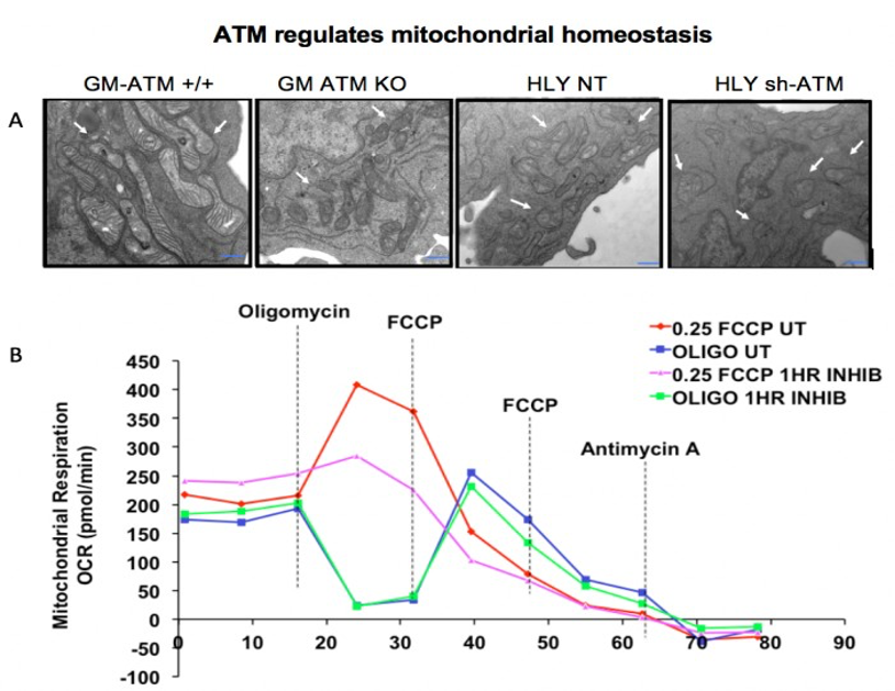
Fig. 2: (A) Representative EM images of mitochondrial structure in non cancer (GM) ATM +/+ & ATM-/- & DLBCL cell lines, HLY-NT & sh-HLY. ATM
unmutated (wt) malignant B-cells consisted of mixed population of tubular and smaller mitochondria compared to typical bacillus-shaped mitochondria in
normal B-cells. Outer membrane was fragmented & a significant lack of the cristae structure within the mitochondria of ATM-/- DLBCL cells. (B)
OCR traces from DLBCL cell line HLY. Cells were chemically inhibited for ATM signaling with KU-55933. Inhibition of ATM
decreased respiration rate in HLY.

Fig. 3: The hyperpolarized 13C signal at ambient temperature after dissolution. Blue and red spectra correspond to metabolic process in the presence of HLY
NT (+/+) and HLY SH (-/-). Both spectra are normalized with respect to maximum pyruvate signal. HLY sh-ATM (-/-) shows higher conversion of pyruvate into
lactate. In the spectra pyruvate, lactate, pyruvate hydrate, alanine and bicarbonate signals are at 172.8, 184.9, 181, 178.4, and 162.7 ppm, respectively.