Chantal Tax1,2, Edwin Versteeg1, Dennis J.W. Klomp1, Martijn F. Froeling1, Alberto de Luca1, and Jeroen C.W. Siero1,3
1University Medical Center Utrecht, Utrecht, Netherlands, 2CUBRIC, Cardiff University, Cardiff, United Kingdom, 3Spinoza Centre for Neuroimaging Amsterdam, Amsterdam, Netherlands
1University Medical Center Utrecht, Utrecht, Netherlands, 2CUBRIC, Cardiff University, Cardiff, United Kingdom, 3Spinoza Centre for Neuroimaging Amsterdam, Amsterdam, Netherlands
This work focuses on the importance of strong gradients - here provided by a gradient head insert - for high SNR and short TE diffusion imaging at 7T. Proof-of-principle images show that a short TE (21 ms) at a b-value of 1000 s/mm2 is achievable using an EPI-readout.

Maximum achievable b-value as a function of TE, for the gradient head insert (solid lines) and body gradients (dashed lines). RF pulse durations of 4 and 6 ms were assumed. The curves are shown for varying readout times [0, 5, 10, 15] ms e.g. depending on resolution and FOV, with a readout time of 0 ms representing a readout strategy starting at the centre of k-space (e.g. spiral imaging5,14).
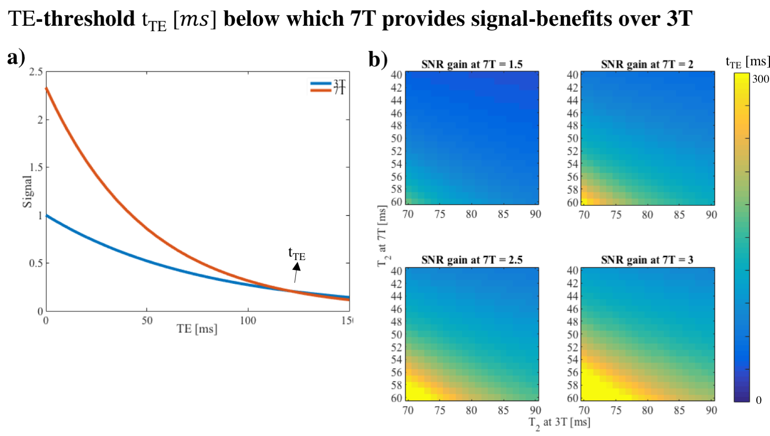
a) Signal decay as a function of TE at 3T (blue) and 7T (red), with tTE the threshold where the signals are equal. A linear increase in signal was assumed as a function of field strength and the apparent T2 was set to 50 and 77 ms at 7T and 3T respectively. b) tTE as a function of other settings for the relative signal gain at 7T - which can depend on e.g. body noise11 - and T2.