Neha A Reddy1,2, Andrew D Vigotsky1,3, Rachael C Stickland2, and Molly G Bright1,2
1Department of Biomedical Engineering, Northwestern University, Evanston, IL, United States, 2Department of Physical Therapy and Human Movement Sciences, Feinberg School of Medicine, Northwestern University, Chicago, IL, United States, 3Department of Statistics, Northwestern University, Evanston, IL, United States
1Department of Biomedical Engineering, Northwestern University, Evanston, IL, United States, 2Department of Physical Therapy and Human Movement Sciences, Feinberg School of Medicine, Northwestern University, Chicago, IL, United States, 3Department of Statistics, Northwestern University, Evanston, IL, United States
Adding an absolute chest position regressor to RETROICOR for physiological noise correction may explain additional variance in spinal cord fMRI data during breath-hold tasks.
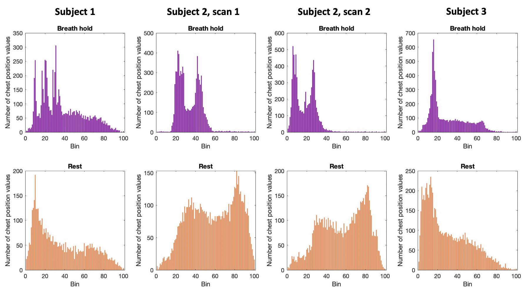
Figure 1. Respiratory cycle histogram created from respiration belt trace of chest position, plotted for each scan. Histograms created from chest position data during breath-hold task scans (top row) and resting-state scans (bottom row). In breath-hold scans, extra peaks at lower bins are due to prolonged low chest position during end-expiratory breath holds. Long tails at higher bins are due to large inhalations during recovery breaths post-breath hold.
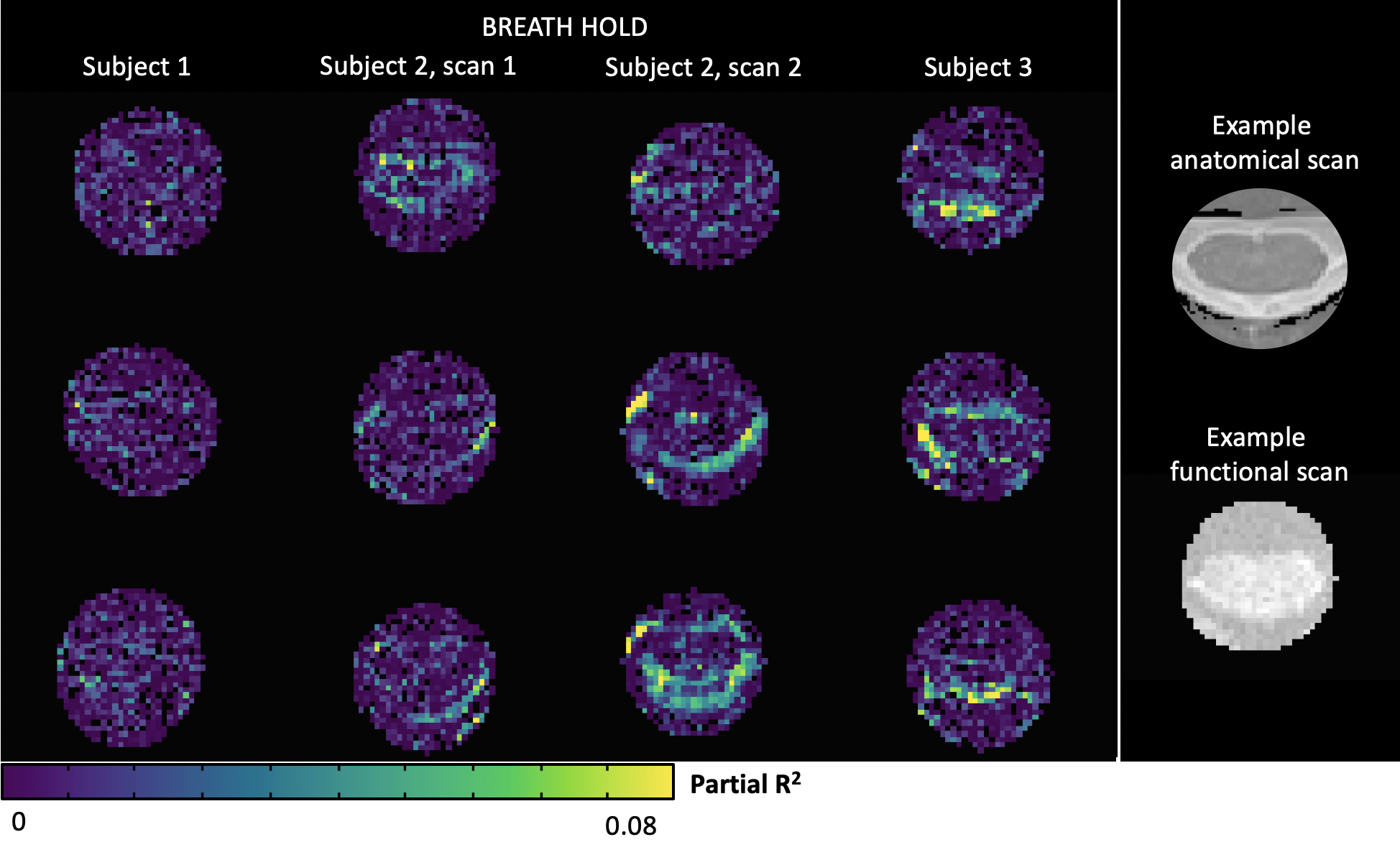
Figure 2. Maps of partial R2 values from adding a chest position regressor compared to RETROICOR alone during breath-hold scans. Three cervical spinal cord transverse slices are shown at approximately the same levels for each subject. Example anatomical and functional scans of the spinal cord are shown in the rightmost column for context.