Merlin M. Weeda1, Alexandra de Sitter1, Iman Brouwer1, Mitchell M. de Boer1, Rick J. van Tuijl1, Petra J.W. Pouwels1, Frederik Barkhof1,2, and Hugo Vrenken1
1Radiology and Nuclear Medicine, Amsterdam UMC - Location VUmc, Amsterdam, Netherlands, 2Institutes of Neurology and Healthcare Engineering UCL, London, United Kingdom
1Radiology and Nuclear Medicine, Amsterdam UMC - Location VUmc, Amsterdam, Netherlands, 2Institutes of Neurology and Healthcare Engineering UCL, London, United Kingdom
This novel, robust, flexible and open-source lesion
simulation tool LESIM enables development of accurate grey matter segmentation or
atrophy measurement software in the presence of white matter lesions in
multiple sclerosis.
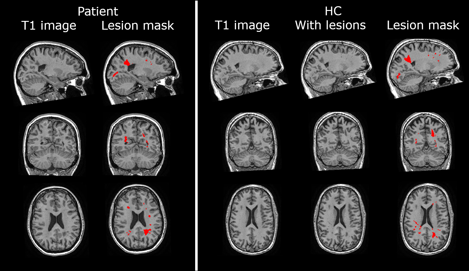
Figure 1. Image of patient and healthy control (HC) with
simulated lesions of patient. Left panel, from left to right: native 3DT1
patient image; and native image with the transformed lesion mask (red). Right
panel, from left to right: native 3DT1 HC image; HC image with simulated
lesions; and HC image with the simulated lesion mask (red).
Note that the lesion mask of the patient was manually
outlined on a FLAIR image and transformed to the T1 image with nearest neighbor
interpolation. GM folds (as visible in top left corner of HC axial slice) are
not segmented as lesions.
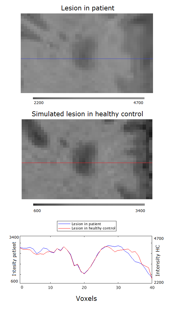
Figure 3. Intensity plot of the intersection of a
patient lesion (blue) and a simulated lesion (red) with surrounding tissue.
Note the intensity axes are different for the patient lesion and simulated
lesion.