Yan Tan1, Wenwei Shi2, Xiaochun Wang1, and Hui Zhang1
1Department of Radiology, First Hospital of Shanxi Medical University, Taiyuan, China, 2Department of Radiology, Zhongda Hospital, Southeast University, Nanjing, China
1Department of Radiology, First Hospital of Shanxi Medical University, Taiyuan, China, 2Department of Radiology, Zhongda Hospital, Southeast University, Nanjing, China
rMK can differentiate
recurrent tumor from pseudoprogression with high diagnostic accuracy, and its
application value is similar to that of rCBV. The combination of rMK and rCBV
improves diagnostic performance compared to either technique alone.

ROC curves of DKI and DSC MRI parameters for differentiating glioma
recurrence from pseudoprogression.
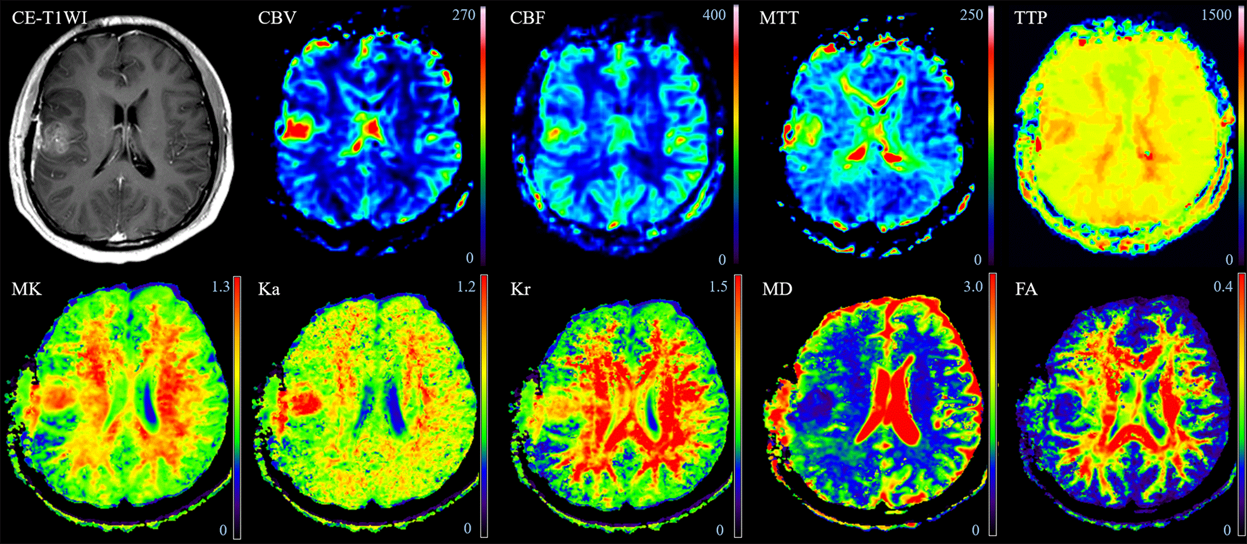
A 21-year-old man with tumor recurrence confirmed by repeat surgery was
initially a high-grade glioma (WHO IV). An increasing
enhanced lesion in the right frontal and temporal lobes was detected on CE-T1WI
with increased CBV, CBF, MTT, TTP, MK, Ka and Kr values, except for MD and FA
values.