Upasana Upadhyay Bharadwaj1, Cynthia T Chin1, Valentina Pedoia1, and Sharmila Majumdar1
1Radiology, University of California, San Francisco, San Francisco, CA, United States
1Radiology, University of California, San Francisco, San Francisco, CA, United States
This study presents classification of facet arthropathy from MRI using zero-shot facet detection followed by binary classification. Our model achieves an AUC of 0.916 with sensitivity and specificity of 97.8% and 64.1%, respectively and can potentially enhance the clinical workflow.
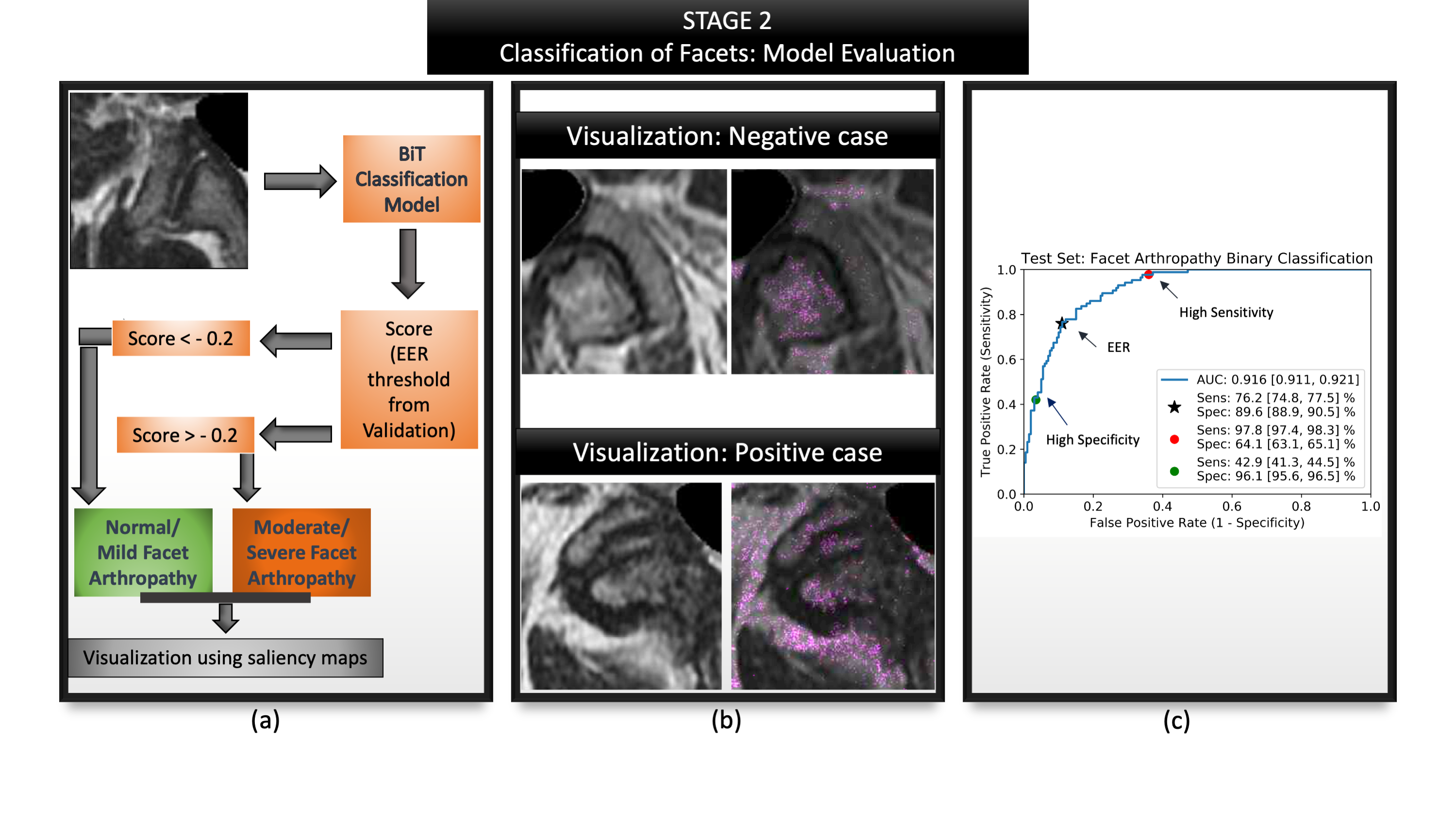
Figure 5: Summarizes the evaluation of second stage: facet classification. (a) Visualizes the entire evaluation pipeline where a patch is passed as input (b) Visualization of the model's predictions via saliency maps shows clinically valuable features being highlighted- image above highlights the superior articular portion of the facet as well as the ligamentum flavum; image below highlights the superior and inferior portions of the facet, synovium; (c) ROC curve highlighting AUC, sensitivity and specificity at various operating points along with their confidence intervals.
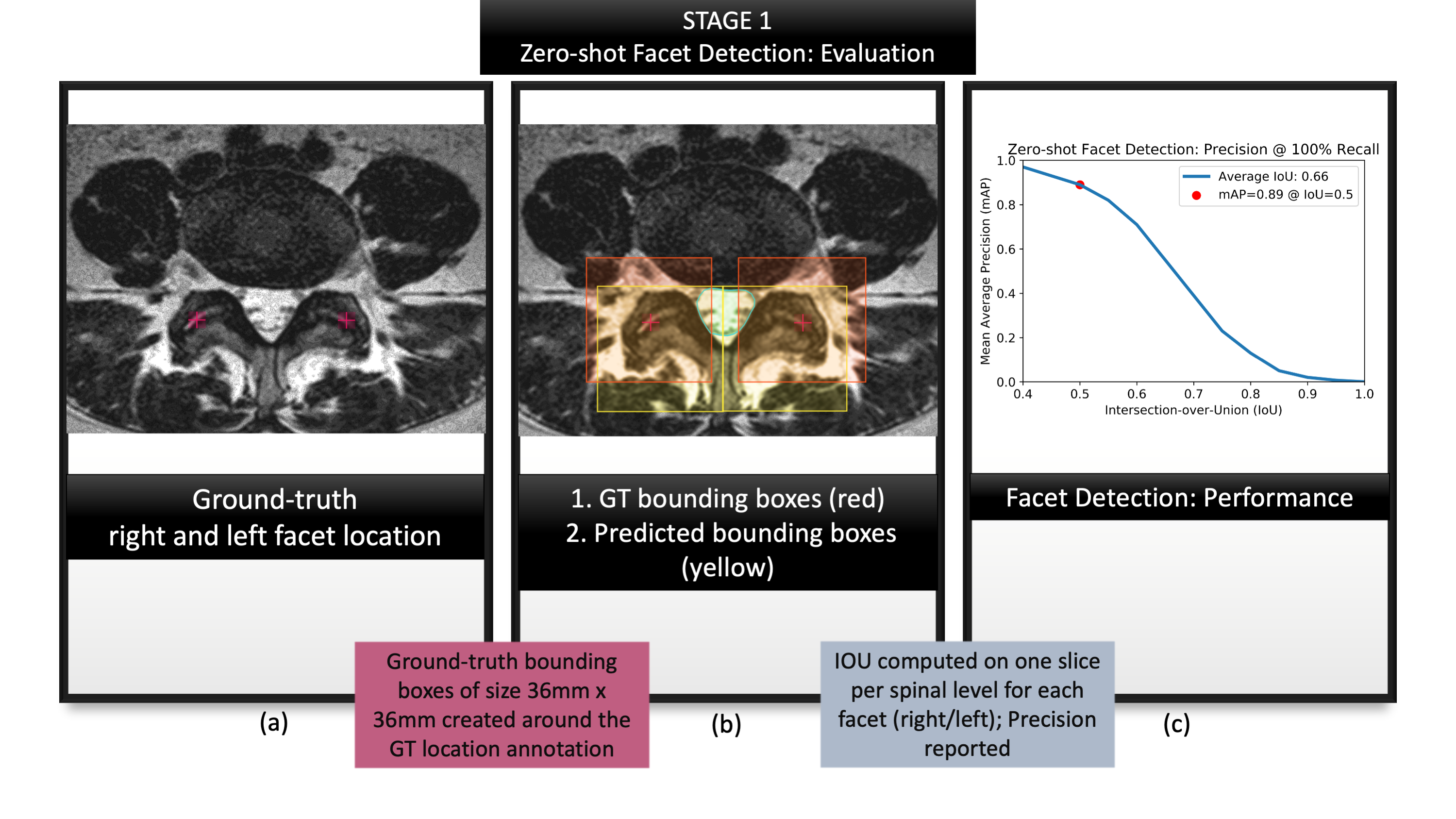
Figure 3: Summarizes the evaluation of first
stage: zero-shot facet detection. (a) visualizes location coordinates annotated
on the T2-w axial slices by a neuroradiologist. These location coordinates were
used purely for evaluating our localization, and not for training our models;
(b) visualizes ground-truth bounding boxes generated from the location
coordinates in (a) shown in red against predicted bounding boxes from zero-shot
detection, shown in yellow.; (c) characterizes the performance with a mAP-IoU
graph.
