Saeed Jerban1, Yajun Ma1, Zhao Wei1, Meghan Shen1, Amir Masoud Afsahi1, Zubiad Ibrahim1, Alecio Lombardi1,2, Douglas G Chang3, Eric Y Chang1,2, and Jiang Du1
1Radiology, University of California, San Digeo, La Jolla, CA, United States, 2Radiology Service, VA San Diego Healthcare System, San Diego, CA, United States, 3Orthopaedic Surgery, University of California, San Digeo, La Jolla, CA, United States
1Radiology, University of California, San Digeo, La Jolla, CA, United States, 2Radiology Service, VA San Diego Healthcare System, San Diego, CA, United States, 3Orthopaedic Surgery, University of California, San Digeo, La Jolla, CA, United States
UTE-MRI-based
estimation of collagen and water proton contents in tibial midshaft were
significantly different between OPo, OPe, and healthy control subjects. UTE-MRI
measures in tibial midshaft were significantly correlated with hip DEXA
T-score.
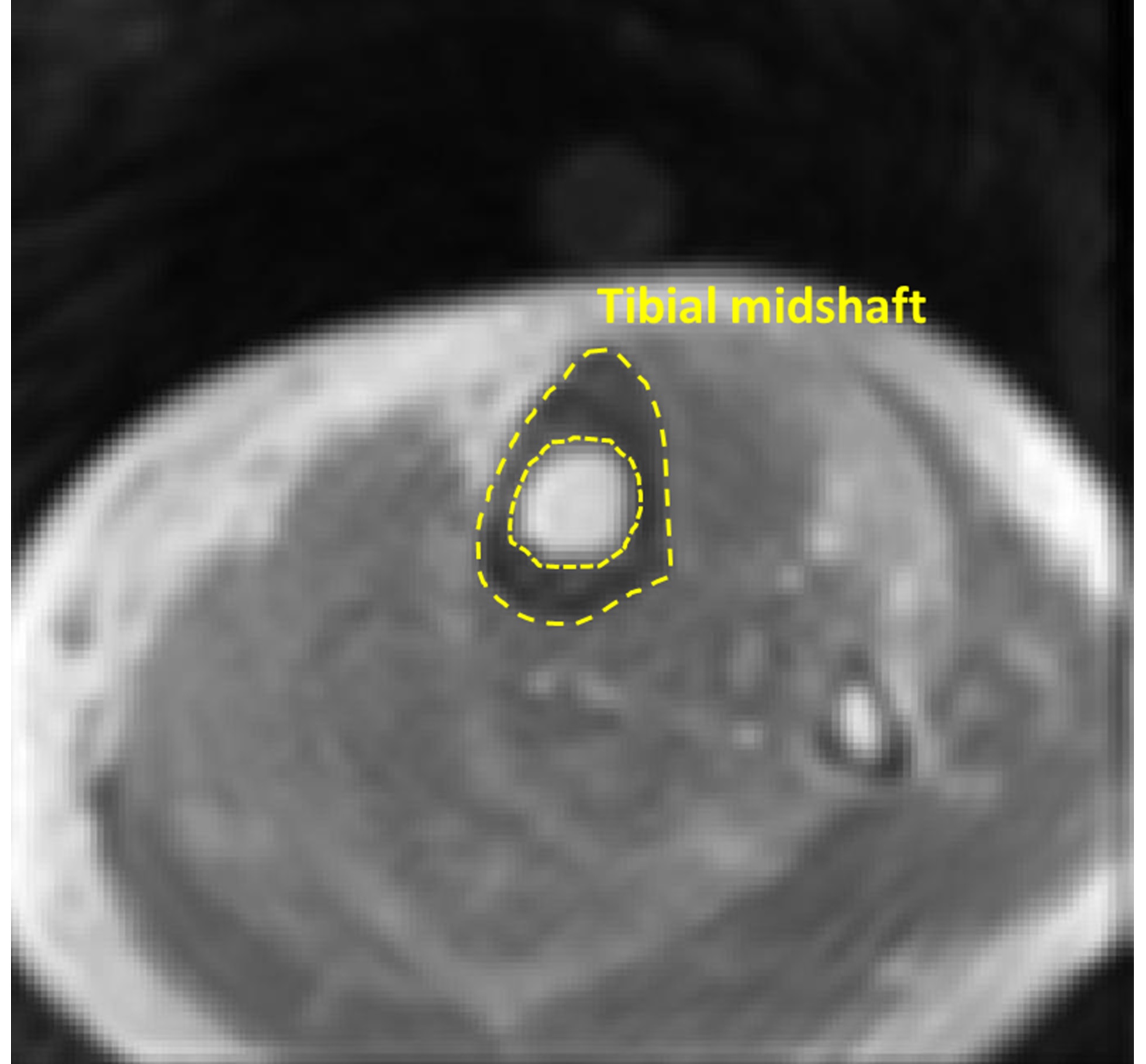
Figure 2: A
representative UTE-Cones-MT MRI image of the lower leg of a 37-year-old female
subject. A representative region of interest is depicted on cortical bone in
tibial midshaft (yellow).
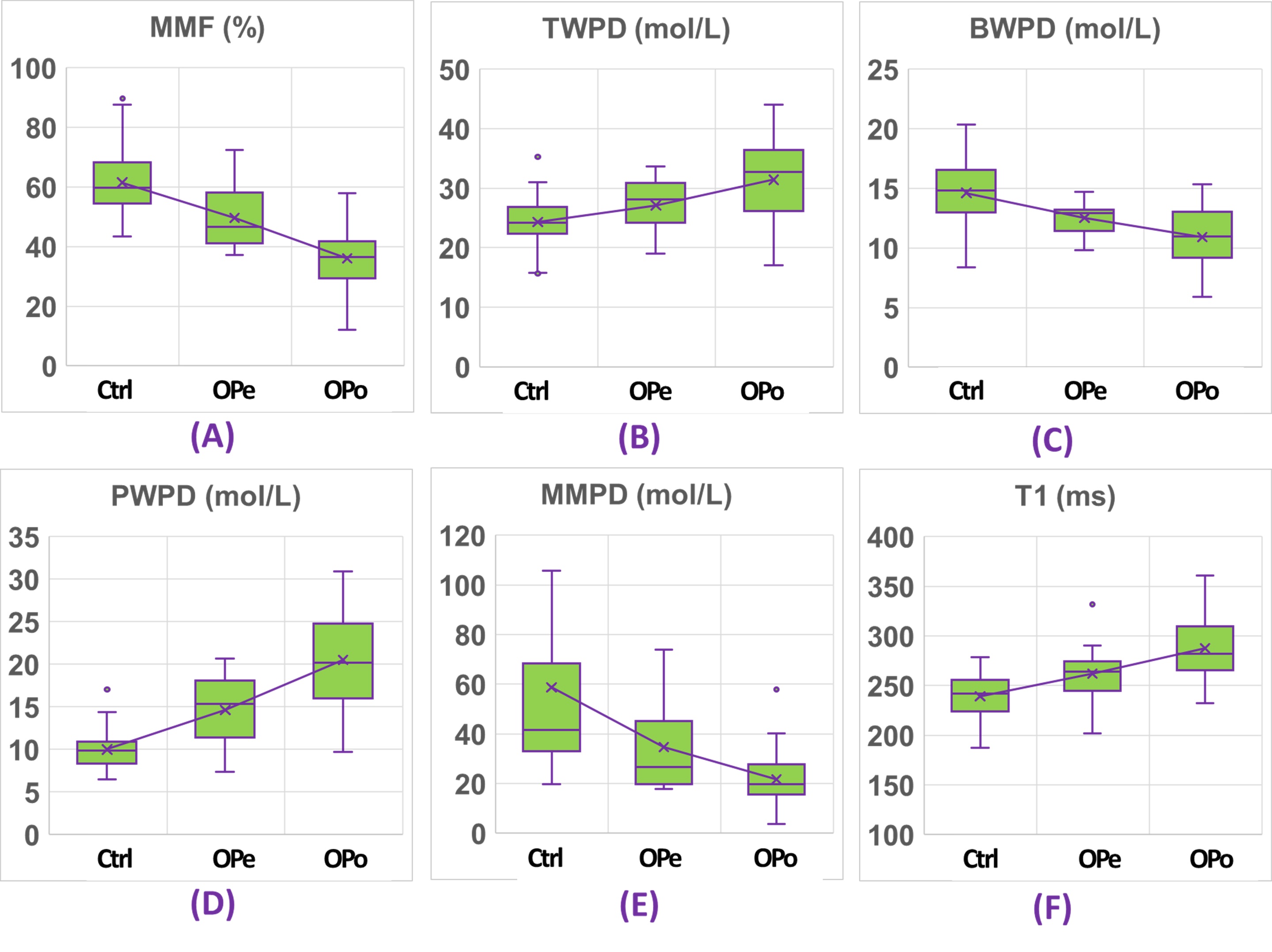
Figure 4: Boxplots of (A) MMF, (B)
TWPD, (C) BWPD, (D) PWPD, (E) MMPD, and (F) T1 values in OPe, OPo, and Ctrl
cohorts. Average, median, SD, first, and third quartiles values are indicated
in the boxplots.