Alecio Lombardi1,2, Jonathan Wong1,2, Rachel High1,2, Ya-Jun Ma2, Adam Searleman2, Saeed Jerban2, Qingbo Tang1,2, Jiang Du1,2, Patrick Frost3,4, and Eric Y. Chang1,2
1Radiology Service, Veterans Affairs San Diego Healthcare System, San Diego, CA, United States, 2Radiology, University of California, San Diego, CA, United States, 3Greater Los Angeles Veteran Administration Healthcare System, Los Angeles, CA, United States, 4University of California, Los Angeles, CA, United States
1Radiology Service, Veterans Affairs San Diego Healthcare System, San Diego, CA, United States, 2Radiology, University of California, San Diego, CA, United States, 3Greater Los Angeles Veteran Administration Healthcare System, Los Angeles, CA, United States, 4University of California, Los Angeles, CA, United States
Multiple myeloma (MM) is a malignant plasma cell disease. Adaptive responses to hypoxia may be an essential element in its progression. The purpose of this study was to determine the feasibility of acidoCEST MRI for pHe measurement on a mouse model of MM with comparison with GLUT1 staining.

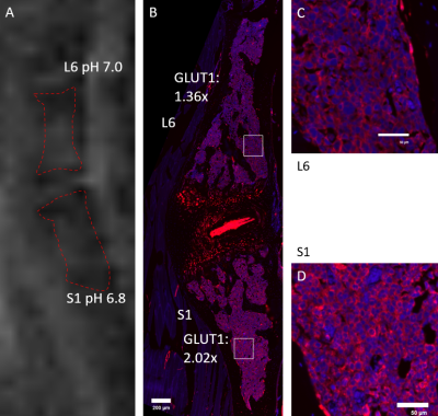
Figure 3. AcidoCEST FISP MRI sequence of mouse's lumbosacral region and correspondent immunofluorescence GLUT1 staining. (A) Average pH measurements of L6 (7.0) and S1 (6.8) were obtained with AcidoCEST. (B, C, D) Correspondent immunofluorescence GLUT1 staining shows higher average GLUT1 staining in S1 (2.02x) compared with L6 (1.36x), inversely correlated with the pH measurements.

Figure 4. AcidoCEST FISP MRI sequence of mouse's coccygeal region and correspondent immunofluorescence GLUT1 staining. (A) Average pH measurements of C1 (6.9) and C2 (6.6) were obtained with AcidoCEST. (B) Immunofluorescence GLUT1 staining shows higher average GLUT1 staining in C2 (1.99x) compared with C1 (1.24x), inversely correlated with the pH measurements.