Yitong Li1, Shuang Hu1, Weiyin Vivian Liu2, and Xiaoming Li1
1Tongji Hospital, Tongji Medical College, Huazhong University of Science and Technology, Wuhan, China, 2MR Research, GE Healthcare, Beijing, China
1Tongji Hospital, Tongji Medical College, Huazhong University of Science and Technology, Wuhan, China, 2MR Research, GE Healthcare, Beijing, China
Zero echo time imaging had higher accuracy and reliability for detection of sacroiliac
joint bone erosions in patients with suspected axial spondyloarthritis,
compared to a routine T1-weighted fast spin echo.
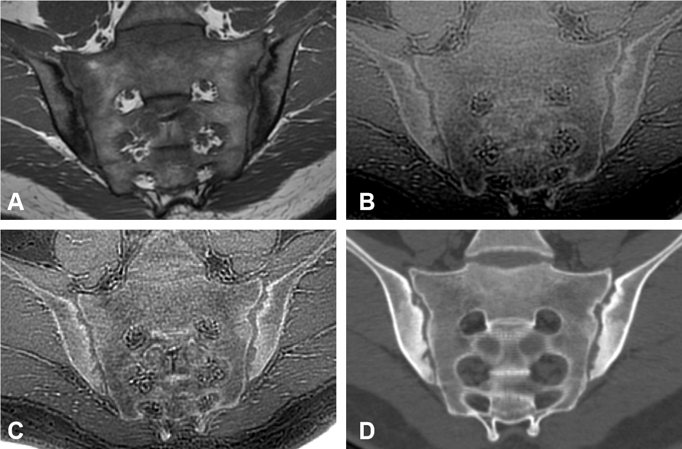
Figure 1: A
25-year-old man with a 2-year history of axSpA. (A) Oblique coronal T1 FSE; (B)
and (C) oblique coronal ZTE without and with postprocessing; (D) Oblique coronal CT. ZTE exhibits similar
bone contrast to CT and provides a similar depiction of erosions in various
areas on CT. ZTE with postprocessing has
higher contrast and sharper and clearer edges. The erosions in the inferior
quadrant of right iliac are not well depicted on the T1 FSE.
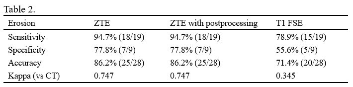
Table 2: Sensitivity, specificity, accuracy and the consistency for
erosion detection of three images, compared to CT.