Hiroaki Takahashi1, Kotaro Yoshida2, Akira Kawashima3, Num Ju Lee4, Adam T Froemming4, Daniel A Adamo4, Ashish Khandelwal4, Candice W Bolan5, Matthew T Heller6, Robert P Hartman4, Bohyun Kim4, Kenneth A Philbrick4, Rickey E Carter5, Lance A Mynderse4, Mitchell R Humphreys6, and Naoki Takahashi4
1Department of Radiology, Mayo Clinic, Rochester, Rochester, MN, United States, 2Department of Diagnostic Radiology, Kanazawa University School of Medical Science, Kanazawa, Japan, 3Department of Radiology, Mayo Clinic, Arizona, Scottsdale, AZ, United States, 4Mayo Clinic, Rochester, Rochester, MN, United States, 5Mayo Clinic, Florida, Jacksonville, FL, United States, 6Mayo Clinic, Arizona, Scottsdale, AZ, United States
1Department of Radiology, Mayo Clinic, Rochester, Rochester, MN, United States, 2Department of Diagnostic Radiology, Kanazawa University School of Medical Science, Kanazawa, Japan, 3Department of Radiology, Mayo Clinic, Arizona, Scottsdale, AZ, United States, 4Mayo Clinic, Rochester, Rochester, MN, United States, 5Mayo Clinic, Florida, Jacksonville, FL, United States, 6Mayo Clinic, Arizona, Scottsdale, AZ, United States
The
optimal method for measuring ADC values for differentiating csPCa and non-csPCa
on prostate MRI are 2D-ROI placed on the lowest ADC area using 6-8 mm2.
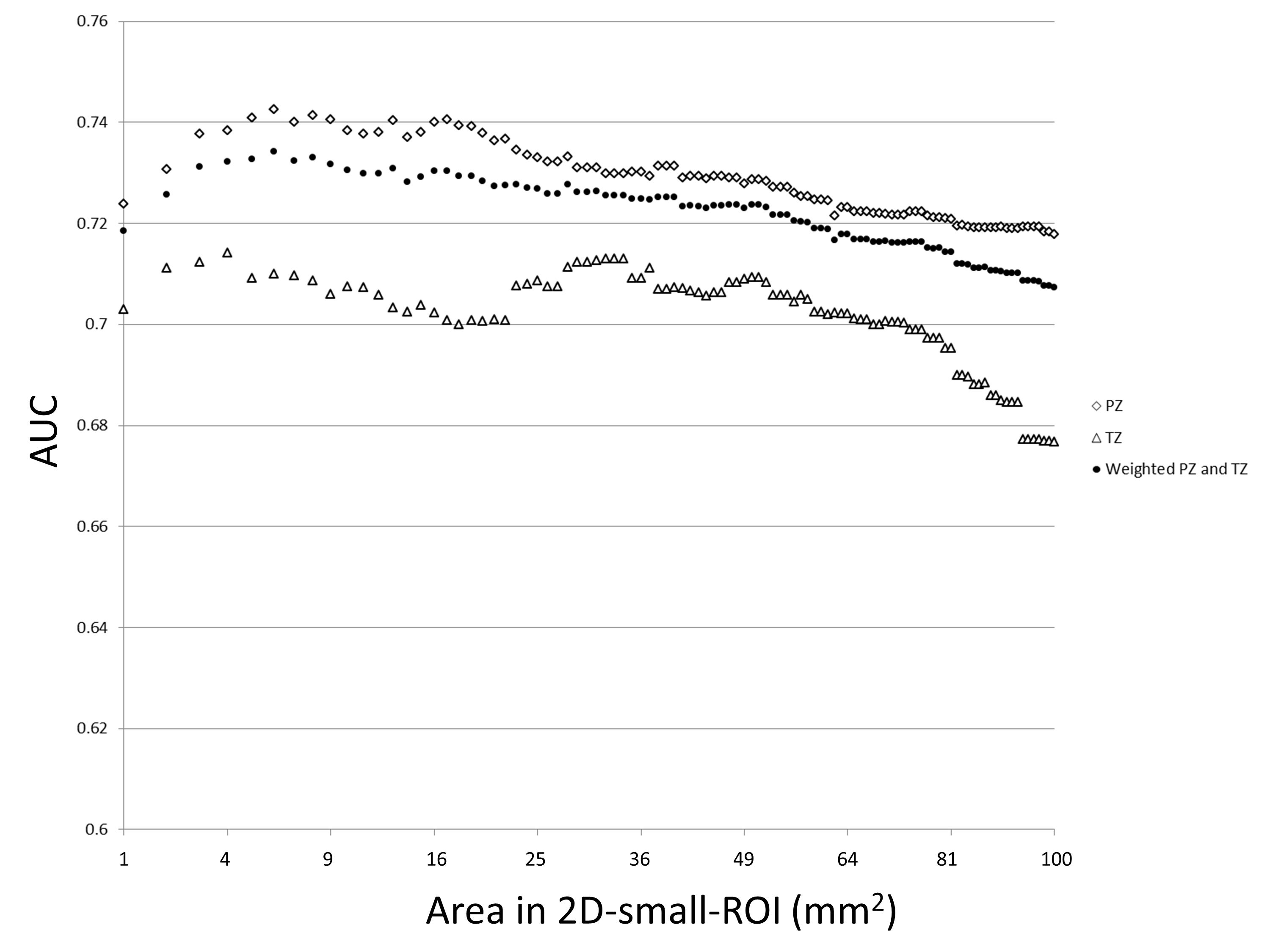
Fig.1. A dot
plot of AUC values of receiver operating characteristics in differentiation of
presence or absence of clinically significant prostate cancer for 2D-small-ROI
with different area
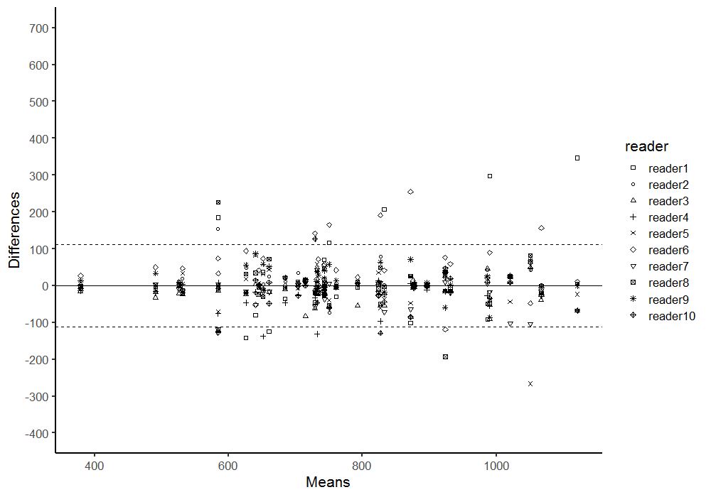
Fig.5. Bland-Altman
plots of ADC values measured by using 3D-whole-lesion-ROI method (10th
percentile). 95% limits of agreement among the readers on was +/-112.