Dharmesh Singh1, Virendra Kumar2, Chandan J Das3, Anup Singh1, and Amit Mehndiratta1
1Centre for Biomedical Engineering (CBME), Indian Institute of Technology (IIT) Delhi, New Delhi, India, 2Department of NMR, All India Institute of Medical Sciences (AIIMS) Delhi, New Delhi, India, 3Department of Radiology, All India Institute of Medical Sciences (AIIMS) Delhi, New Delhi, India
1Centre for Biomedical Engineering (CBME), Indian Institute of Technology (IIT) Delhi, New Delhi, India, 2Department of NMR, All India Institute of Medical Sciences (AIIMS) Delhi, New Delhi, India, 3Department of Radiology, All India Institute of Medical Sciences (AIIMS) Delhi, New Delhi, India
Texture analysis based machine learning approaches are presented in characterization of PI-RADS v2 grades of prostate cancer using T2WI. The
use of texture features extracted from T2WI, DWI and ADC improve the
accuracy of prostate cancer characterization by almost 23% compared to T2WI alone
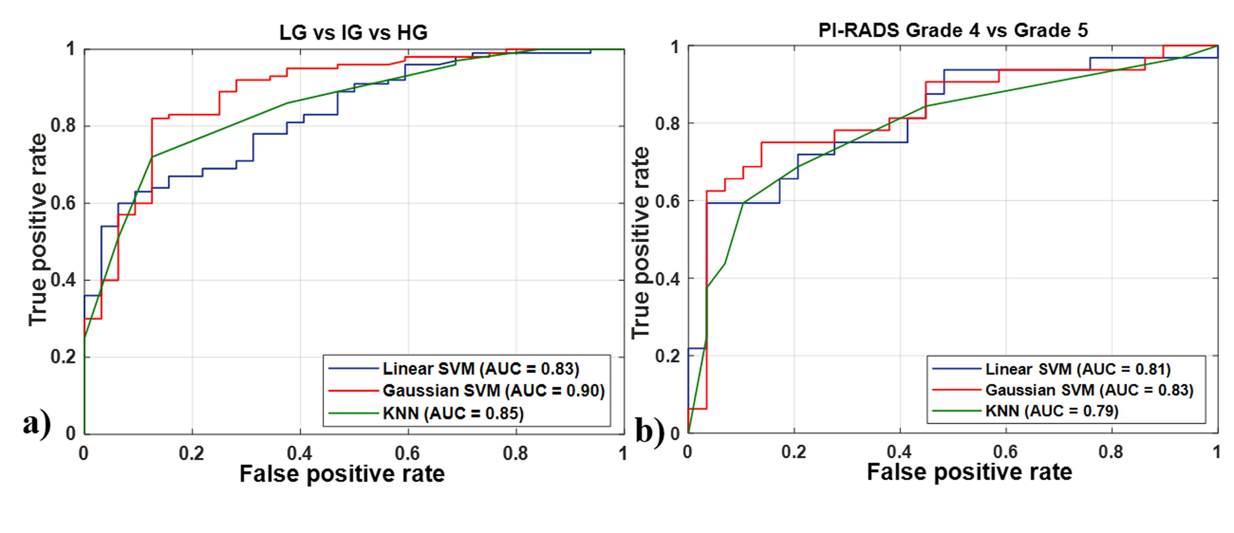
Figure 1: ROC plot for a) LG vs. IG vs. HG and
b) PI-RADS grade 4 vs. grade 5 classification
using T2WI. Red curves stand for the performance of
linear SVM, green curves for Gaussian SVM and blue curves for KNN classifier. ROC = Receiver-operating
characteristics, CV = cross-validation, SVM = Support vector machine, KNN =
K-nearest neighbour, DWI = Diffusion-weighted imaging, ADC = Apparent diffusion
coefficient
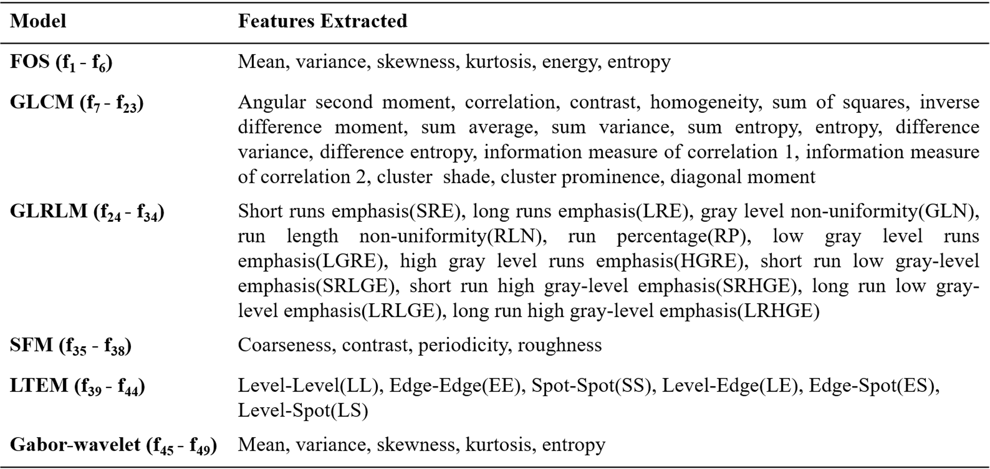
Table
1:
Features extracted from different
texture models. FOS = First order statistics, GLCM = Gray level
co-occurrence matrix, GLRLM = Gray level run length matrix, SFM = Statistical
feature matrix, LTEM = Law's texture energy measures