Seb Harrevelt1, Lieke Wildenberg2, Dennis Klomp2, C.A.T. van den Berg2, Josien Pluim3, and Alexander Raaijmakers1
1TU Eindhoven, Utrecht, Netherlands, 2UMC Utrecht, Utrecht, Netherlands, 3TU Eindhoven, Rossum, Netherlands
1TU Eindhoven, Utrecht, Netherlands, 2UMC Utrecht, Utrecht, Netherlands, 3TU Eindhoven, Rossum, Netherlands
We find that a deep learning approach to bias field suppression in 7T images can achieve good results, even when trained on artificially created 7T data that is based on 1.5T data.
Applying
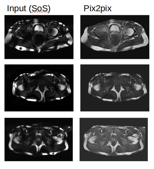
the pix2pix model to real measured 7T MRI data over different
anatomies (left)
and its corresponding output (right).
Note
that instead of showing the model input (8 single-channel images),
the figure shows the sum of magnitude of these input images.

Graphical
representation of data creation pipeline. Going from left to right,
we start with a set of simulated B1+ and B1- fields, and a target
prostate image. The B1+ and B1- fields are being registered to the
shape of the prostate data whereafter the registered B1+ data is
shimmed and scaled to mimic the signal of a T2w acquisition. Now
combining the scaled B1+, B1- and prostate image results in the
8-channel input data for our model.