Sivakami Avadiappan1, Jeffrey Nelson2, Marc Mabray3, Blaine Hart3, Leslie Morrison4, Atif Zafar4, Michel Torbey4, Helen Kim2,5, and Janine Lupo1
1Department of Radiology and Biomedical Imaging, University of California San Francisco, San Francisco, CA, United States, 2Department of Anesthesia and Perioperative Care, University of California San Francisco, San Francisco, CA, United States, 3Department of Radiology, University of New Mexico, Albuquerque, NM, United States, 4Department of Neurology, University of New Mexico, Albuquerque, NM, United States, 5Department of Epidemiology and Biostatistics, University of California San Francisco, San Francisco, CA, United States
1Department of Radiology and Biomedical Imaging, University of California San Francisco, San Francisco, CA, United States, 2Department of Anesthesia and Perioperative Care, University of California San Francisco, San Francisco, CA, United States, 3Department of Radiology, University of New Mexico, Albuquerque, NM, United States, 4Department of Neurology, University of New Mexico, Albuquerque, NM, United States, 5Department of Epidemiology and Biostatistics, University of California San Francisco, San Francisco, CA, United States
Lesion burden and count of segmented cerebral cavernous malformation lesions were significantly correlated with age and obesity. Lesion burden in the brainstem and temporal lobes were related to hemorrhagic and seizure events.
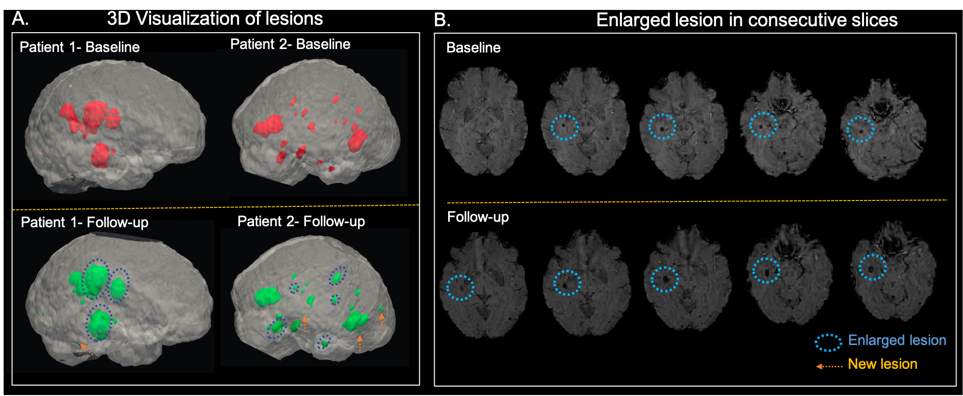
Figure 2. A. 3D visualization of CCM lesions in baseline (pink) and follow-up (green) scans for 2 patients. The lesions that are enlarged in the follow-up scan are highlighted with the blue circle while new lesions are denoted by an arrow. B. The enlargement of the lesion (blue circle) in the follow-up scan compared to the baseline is evident in the consecutive slices from a patient.
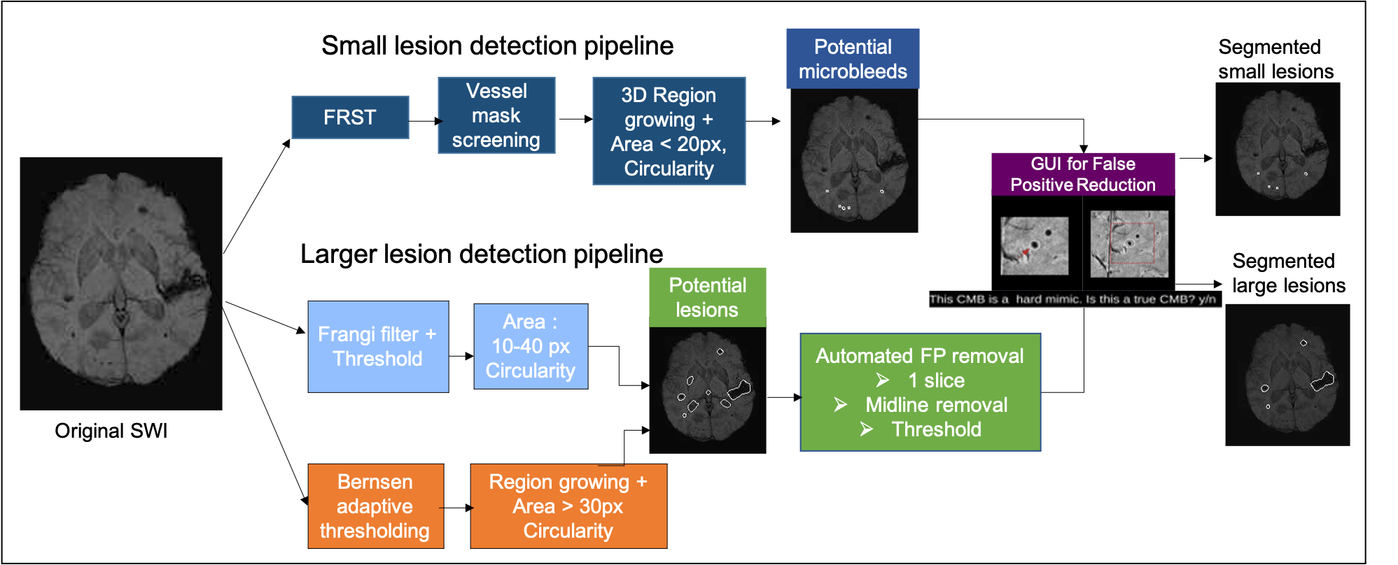
Figure 1. Pipeline for detecting the different sized lesions. (Top) Pipeline for small lesion detection. (Bottom) Pipeline for larger lesion detection. The potential lesion candidates are fed to the GUI where the user labels the lesions.