Hui Wang1, Xianchang Zhang2, Quanzhi Feng1, Yutian Li1, Jinli Li1, Yujun Wang3, Guangzhao Yang3, Qingle Kong2, Zihao Zhang4, and Tong Han1
1Radiology, Tianjin Huanhu Hospital, Tianjin, China, 2MR Collaboration, Siemens Healthcare Lid., Beijing, China, 3Radiology, Tongde Hospital of Zhejiang Province, Hangzhou, China, 4State Key Laboratory of Brain and Cognitive Science, Institute of Biophysics, Chinese Academy of Sciences, Beijing, China
1Radiology, Tianjin Huanhu Hospital, Tianjin, China, 2MR Collaboration, Siemens Healthcare Lid., Beijing, China, 3Radiology, Tongde Hospital of Zhejiang Province, Hangzhou, China, 4State Key Laboratory of Brain and Cognitive Science, Institute of Biophysics, Chinese Academy of Sciences, Beijing, China
Non-stenotic MCA plaques are
effectively detected using HR-VWI. Coupled with a derived biomarker of
laterality index, this method may be promising for the identification of BAD in SSIs.
[A1]is? might be?
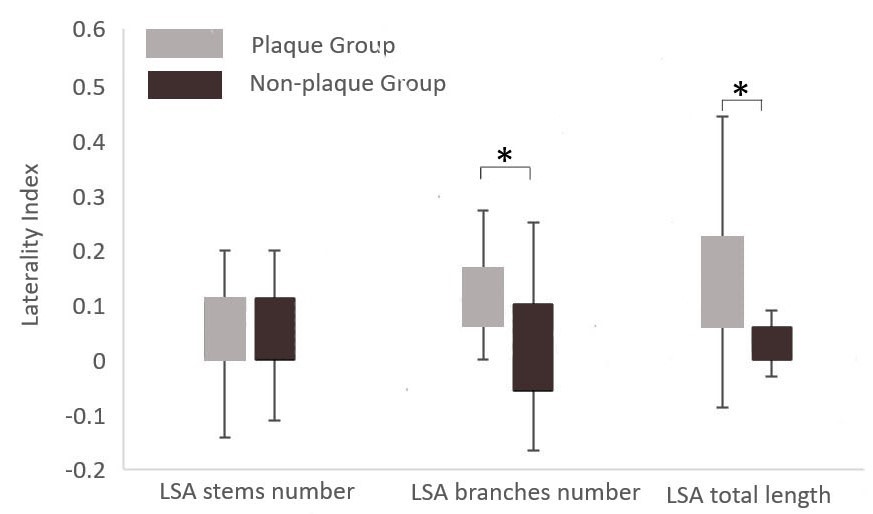
Figure 3: Comparison of laterality index
(L_index) of LSA morphological parameters between the plaque and non-plaque
groups. The L_Index of LSA branches and total length in the plaque group were significantly
higher (*, p<0.05) than those in the non-plaque. There was no statistical
difference in L_Index of LSA stems.
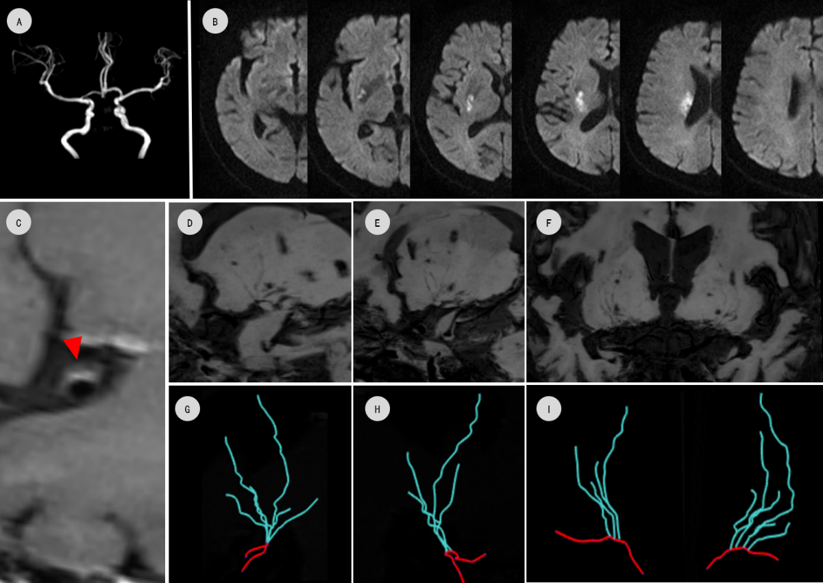
Figure1: Plaque group, a 69-year-old female
with right lenticulostriate infarction (B) and non-stenotic MCA (A). C: HR-VWI
shows the plaque on the upper wall of the MCA-M1 segment (red arrow). LSA skeletons
(G-I, from D-E) are shown. The numbers of LSA stems and branches and the total
length of LSA on infarcted and healthy sides were 4/6/125 mm and 3/4/89 mm, respectively.
The calculated laterality indexes were 0.14/0.30/0.17, respectively.