Jackson Moore1, Maria Aristova1, Ramez Abdalla1, Ann Ragin1, Eric Russell1, Fan Caprio2, Michael Hurley1, Susanne Schnell3, Sameer A. Ansari1, and Michael Markl1
1Radiology, Northwestern University, Chicago, IL, United States, 2Neurology, Northwestern University, Chicago, IL, United States, 3Universitaet Greifswald, Greifswald, Germany
1Radiology, Northwestern University, Chicago, IL, United States, 2Neurology, Northwestern University, Chicago, IL, United States, 3Universitaet Greifswald, Greifswald, Germany
Cerebrovascular
dual-venc 4D flow MRI: Comprehensive assessment of arterial pulsatility and resistance
measures in intracranial atherosclerotic disease
patients
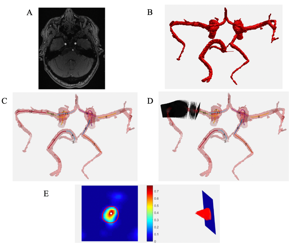
Figure 1: Analysis workflow. Preprocessing
(noise, phase offset, velocity anti-aliasing corrections) and generation of
dual-venc phase-contrast MR angiogram workflow not shown. A – TOF MRA DICOMS imported for segmentation. B – 3D segmentation of Circle of Willis from in-house analysis
tool. TOF is registered with PC MRA to extract 4D flow information. C – Vessel branch centerline extraction
for further analysis. D – Placement
of analysis planes along example vessel every 1 mm. E – View of single plane ROI delineating vessel contours, velocity map, and velocity vector profile.
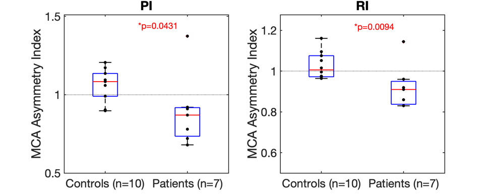
Figure 2: Asymmetry
index analysis results for PI and RI in ICAD patients with symptomatic MCA
vessels. PI and RI comparisons to controls showed statistical significance (p
< 0.05).