Zhangyan yang1,2, Feng Wang1,3, Chaohui Tang1, Li Min Chen1,3, and Gore C. John1,2,3
1Institute of Imaging Science, Vanderbilt University, Nashiville, TN, United States, 2Biomedical Engineering, Vanderbilt University, Nashiville, TN, United States, 3Department of Radiology and Radiological Science, Vanderbilt University, Nashiville, TN, United States
1Institute of Imaging Science, Vanderbilt University, Nashiville, TN, United States, 2Biomedical Engineering, Vanderbilt University, Nashiville, TN, United States, 3Department of Radiology and Radiological Science, Vanderbilt University, Nashiville, TN, United States
High resolution maps of rCBV provide information for studies of brain function and changes in brain. In this study, by using a contrast agent in non-human primates, we quantified rCBV maps, identified alterations of rCBV across regions, and created an rCBV atlas using a brain template.

Figure 1. Registration pipeline of VALiDATe29 squirrel monkey atlas labels (template space) to structural images before MION (subject space). T2* weighted image before MION administration were first skull stripped and down sampled to template size. Then the template image was transformed into subject space by performing affine linear automatic image registration (AIR) followed by non-linear LDDMM registration. Inverse transformation matrices were subsequently applied to the template labels. Labels are transformed to subject space and checked visually using MRICroN.
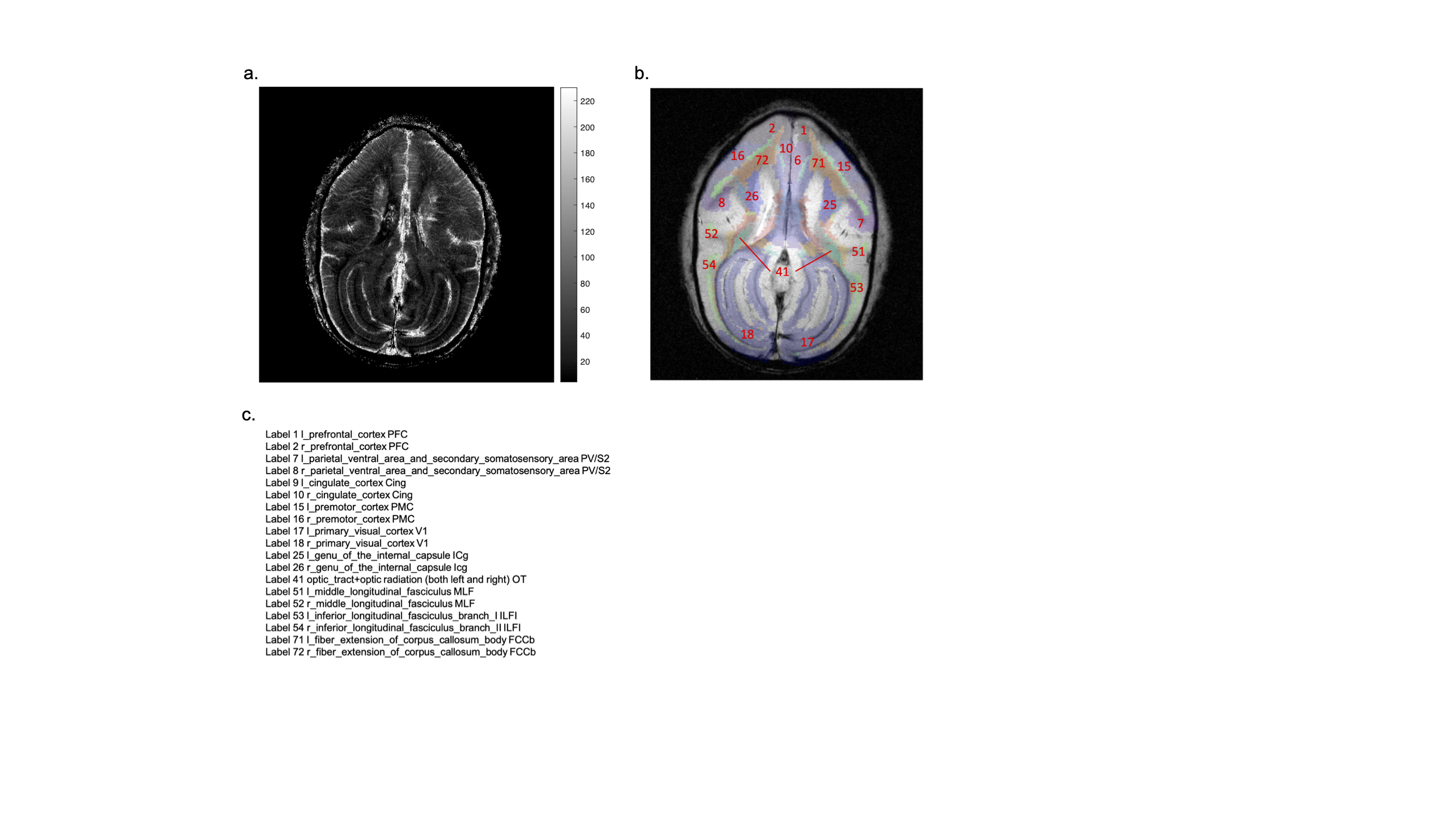
Figure 2. (a) Characteristic rCBV map of one subject. The 13th slice is shown. (b) structural T2* image before MION overlayed with transformed labels in subject space for the same slice. Structural image is in gray scale and labels are indicated in different color. (c) label names, the label numbers are consistent with numbers in b.