Kristina Mary Pelkola1,2, Onur Afacan1,2, Tess E. Wallace1,2, Pauline Connaughton1, Jenna McKay1, Joseph Zmuda1, Camilo Jaimes1, and Simon K. Warfield1,2
1Radiology, Boston Children's Hospital, Boston, MA, United States, 2Computational Radiology Laboratory, Boston Children's Hospital, Boston, MA, United States
1Radiology, Boston Children's Hospital, Boston, MA, United States, 2Computational Radiology Laboratory, Boston Children's Hospital, Boston, MA, United States
Magnetic Resonance Imaging (MRI) can be challenging for pediatric
patients due to factors such as the large tunnel, imaging coil, and loud
gradient noises. This can spark anxiety and fear causing them to be uncooperative
and unable to hold still [1,2]. Due to these barriers, pediatric MRI exams can
be plagued with motion artifacts which result in poor diagnostic quality of the
images and make it challenging for Radiologists to interpret [3]. It is a
common practice to administer sedation or anesthesia to attempt to acquire
diagnostic images of pediatric patients. These methods are not only costly and
potentially harmful, but do not eliminate motion artifacts [4]. Exams performed
under sedation and anesthesia can still be affected from breathing and/or
uncontrollable muscle spasms. There is an unmet need for alternative methods to
enable diagnostic imaging exams in the presence of motion.
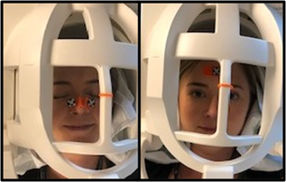
Figure 2: Adult volunteer displaying the
double winged marker, on the left, and the single flat marker, on the right in
a Siemens 64-channel head coil [7].