Weekend Course
MR Physics: From Spins to Scanner II
ISMRM & SMRT Annual Meeting • 15-20 May 2021

| Concurrent 3 | 13:45 - 14:30 | Moderators: Nicolas Boulant & Priti Balchandani |
 |
Simulating Pulses & Sequences
Maxim Zaitsev
This teaching presentation considers basic properties of the Bloch equation along with the practical and efficient methods of solving it. It shows how Bloch equation solvers for arbitrary drive fields can be converted into a core of an MR simulator. Several approaches to building such simulators are discussed along with the brief review of major open-source software packages implementing such functionality.
|
|
| Contrast-topia: Flow & Diffusion
Jennifer McNab
|
||
 |
Contrast-topia: Spectroscopy & Chemical Exchange
Anke Henning, Eleni Demetriou
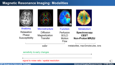
This educational presentation introduces the basic physical principles of magnetic resonance spectroscopy (MRS) / magnetic resonance spectroscopic imaging (MRSI) and Chemical Exchange Saturation Transfer (CEST) imaging. The influence of the nucleus and chemical shift on the resonance frequency is discussed and J-coupling, Nuclear Overhauser Effect and Chemical Exchange are introduced. Confounding effects that need to be calibrated out to yield quantitative results are mentioned. Both methods are compared with each other with respect to their sensitivity and specificity. In addition, the metabolic information that can be extracted from MRS/MRSI and CEST is discussed and clinical and research applications are introduced.
|
|
 |
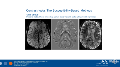
Contrast-topia: The Susceptibility-Based Methods
Sina Straub
Magnetic susceptibility in biological tissue is discussed and its influences in gradient echo imaging. Different imaging methods are explained to exploit tissue susceptibility differences as well as the use of blood-oxygen-level-dependent (BOLD) effect in fMRI and the use of paramagnetic contrast agents for dynamic susceptibility contrast perfusion imaging. Benefits of the use of ultra-high field MRI are highlighted and the choice of imaging parameters is briefly discussed.
|
|
 |
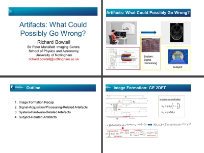
Artifacts: What Could Possibly Go Wrong?
Richard Bowtell
There are many different types of artefacts in MRI – way too many to cover sensibly in a single educational talk – this presentation will therefore focus on a sub-set of artefacts and provide an explanation of the origin of each and introduce ways they can be ameliorated. Example artefact images will be shown, along with the results of simulations that allow the effects of varying imaging parameters on the artefact properties to be probed. We will consider artefacts resulting from: (i) errors in signal acquisition or processing; (ii) system hardware imperfections and (iii) the human subject of the scanning.
|
|
| Putting It All Together: The Scanner
Thomas Foo
There are many considerations when designing a scanner. The purpose, clinical imaging needs, target performance, and complexity of the problem all need to be balanced, with trade-offs made along the way. We will be putting it all together and look at assembling a brain MRI scanner as an example.
|
The International Society for Magnetic Resonance in Medicine is accredited by the Accreditation Council for Continuing Medical Education to provide continuing medical education for physicians.