Digital Poster
Female Pelvis I: Placenta & Pelvic Anatomy
Joint Annual Meeting ISMRM-ESMRMB & ISMRT 31st Annual Meeting • 07-12 May 2022 • London, UK

| Computer # | ||||
|---|---|---|---|---|
0811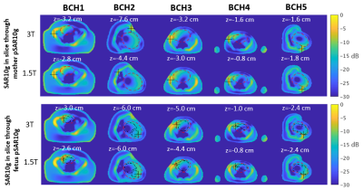 |
36 | Comparison of local SAR and temperature increase between 1.5T and 3T in fetal MRI across five numerical pregnant body models
Filiz Yetisir1, Esra Abaci Turk1,2, Judy A Estroff3,4, Carol Barnewolt3,4, Elfar Adalsteinsson5,6,7, Patricia Ellen Grant1,2,3, and Lawrence L Wald6,8,9
1Fetal-Neonatal Neuroimaging and Developmental Science Center, Boston Children's Hospital, Boston, MA, United States, 2Department of Pediatrics, Boston Children's Hospital, Boston, MA, United States, 3Department of Radiology, Boston Children's Hospital, Boston, MA, United States, 4Maternal Fetal Care Center, Boston Children's Hospital, Boston, MA, United States, 5Department of Electrical Engineering and Computer Science, Massachusetts Institute of Technology, Cambridge, MA, United States, 6Harvard-MIT Division of Health Sciences and Technology, Massachusetts Institute of Technology, Cambridge, MA, United States, 7Institute for Medical Engineering and Science, Massachusetts Institute of Technology, Cambridge, MA, United States, 8Athinoula A. Martinos Center for Biomedical Imaging, Massachusetts General Hospital, Charlestown, MA, United States, 9Department of Radiology, Massachusetts General Hospital, Boston, MA, United States
3T can improve fetal MRI compared to 1.5T, but concerns over increased RF safety risk for the fetus exist due to higher field nonuniformity. Previous studies comparing fetal SAR between two field strengths used either a single pregnant body model or artificial pregnant body models. In this study, we compare SAR and temperature increase in the fetus and mother at 1.5T and 3T using 5 anatomically realistic and diverse pregnant body models. Across these models, we find similar or lower levels of fetal SAR and temperature increase at 3T compared to 1.5T.
|
||
0812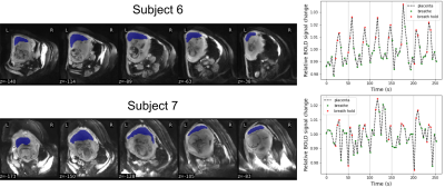 |
37 | Placental MRI: The effect of maternal breath hold on T2* quantified by multi-echo gradient echo EPI
Jeffrey N Stout1, Esra Abaci Turk1, Borjan Gagoski1, Mary Katherine Manhard2,3, William H Barth, Jr.4, Elfar Adalsteinsson5,6, and P. Ellen Grant1
1Fetal-Neonatal Neuroimaging Developmental Science Center, Boston Children's Hospital, Boston, MA, United States, 2Department of Radiology, Cincinnati Children's Hospital Medical Center, Cincinnati, OH, United States, 3Department of Radiology, University of Cincinnati College of Medicine, Cincinnati, OH, United States, 4Maternal-Fetal Medicine, Massachusetts General Hospital, Boston, MA, United States, 5Institute of Medical Engineering and Science, Massachusetts Institute of Technology, Cambridge, MA, United States, 6Electrical Engineering and Computer Science, Massachusetts Institute of Technology, Cambridge, MA, United States
T2* imaging has been the focus of research into the non-invasive MRI assessment of placental function, yet the fundamental physiology that determines placental T2* is under active investigation. We explored the effect of maternal breath holding on placental T2* quantified with multi-echo GRE EPI. We quantified T2* in free-breathing and breath hold periods by identifying voxels that responded to the breath hold stimulus through fitting with a GLM, and found a consistent positive increase in T2* during breath hold. This increase may be evidence that fetal SO2 rises during mild maternal hypercapnia, as has been observed previously in instrumented animals.
|
||
0813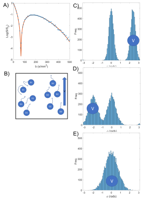 |
38 | Coherent flows detected in placental diffusion weighted data
George Hutchinson1, Neele Dellschaft1, Nia Jones2, Lopa Leach3, and Penny Gowland1
1Sir Peter Mansfield Imaging Centre, The University of Nottingham, Nottingham, United Kingdom, 2Department of Child Health, Obstetrics and Gynaecology, School of Medicine, The University of Nottingham, Nottingham, United Kingdom, 3School of Life Sciences, The University of Nottingham, Nottingham, United Kingdom
Diffusion weighted imaging is commonly used in the placenta to characterise regions of differing structure and flow, including differences between the healthy and compromised placenta. Typically placental diffusion data is described by a combination of exponential decays with increasing diffusion weighting, but we have observed signals that show oscillations in signal or even increasing initial signal and are likely to relate to refocusing caused by coherent flows. By altering our diffusion fitting we can access information about flow patterns and provide more information regarding placental haemodynamics.
|
||
0814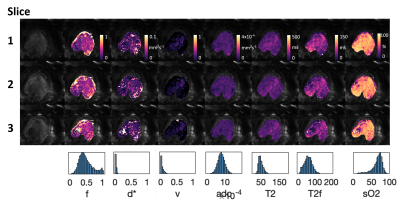 |
39 | Multi-modal MRI reveals changes in placental function following preterm premature rupture of membranes
Andrew Melbourne1, Carla Avena Zampieri1, Dimitra Flouri1, Joseph V Hajnal1, Anna David2, Mary A Rutherford1, Jana Hutter3, and Lisa Story4
1Centre for Medical Engineering, King's College London, London, United Kingdom, 2University College London, London, United Kingdom, 3King's College London, London, United Kingdom, 4Women's Health, King's College London, London, United Kingdom
Preterm premature rupture of membranes (PPROM) complicates up to 40% of deliveries less than 37 completed weeks gestation. Absence of clinical symptoms means that fetal infection can be missed and later complications are significantly higher. There is thus a real need for non-invasive antenatal assessment of the early signs of placental inflammation. This study combines an efficient multimodal Diffusion-Relaxation acquisition with dedicated analysis to study the placental phenotype associated with PPROM. Initial results indicate that multi-modal MRI measurement of placental function is linked to eventual gestational age at delivery.
|
||
0815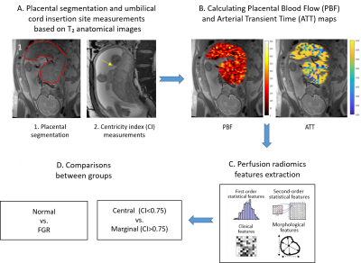 |
40 | Structural-functional interplay of the human placenta: in-vivo study using Pseudocontinuous Arterial Spin Labeling
Daphna Link1, Netanell Avisdris1,2, Xingfeng Shao3, Liat Ben Sira4,5, Liran Hiersch6, Ilan Gull7, Danny J.J. Wang8, and Dafna Ben Bashat1,4
1Sagol Brain Institute, Tel Aviv Sourasky Medical Center, Tel Aviv, Israel, 2School of Computer Science and Engineering, The Hebrew University of Jerusalem, Jerusalem, Israel, 3Laboratory of FMRI Technology (LOFT), USC Stevens Neuroimaging and Informatics Institute, Keck School of Medicine, University of Southern California, Los Angeles, CA, United States, 4Sackler Faculty of Medicine and Sagol School of Neuroscience, Tel Aviv University, Tel Aviv, Israel, 5Division of Pediatric Radiology, Tel Aviv Sourasky Medical Center, Tel Aviv, Israel, 6Division of Obstetrics and Gynecology, Tel Aviv Sourasky Medical Center, Tel Aviv, Israel, 7Ultrasound Unit, Lis Maternity Hospital, Tel Aviv Sourasky Medical Center, Tel Aviv, Israel, 83Laboratory of FMRI Technology (LOFT), USC Stevens Neuroimaging and Informatics Institute, Keck School of Medicine, University of Southern California, Los Angeles, CA, United States
Adequate placental structure and function are crucial for fetal and maternal health. Here we assessed late-gestation placental perfusion (N=50, GA=31-38 weeks) using pseudocontinuous arterial-spin-labeling and radiomics feature-extraction, and their association with umbilical-cord insertion-site and the risk of fetal-growth-restriction (FGR). Increased blood-flow and arterial-transit-time were detected in our normal cases compared to earlier gestation as reported previously. Perfusion features differences were detected in placentas with marginal compared to central cord-insertion, indicating higher intensity heterogeneity. No differences were found between normal and FGR placentas. This study provides normal late-gestation placental perfusion values based on a large cohort, and implies on structural-functional associations.
|
||
0816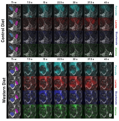 |
41 | Effects of Life-Long Maternal Western Diet on Guinea Pig Placental Metabolism Across Gestation using [1-13C]pyruvate MRI Video Permission Withheld
Lindsay E. Morris1, Lanette J. Friesen-Waldner1, Trevor P. Wade1, Lauren M. Smith2, Mary-Ellen ET. Empey1, Timothy RH. Regnault3,4,5, and Charles A. McKenzie1,5
1Medical Biophysics, Western University, London, ON, Canada, 2Radiation Oncology, University of California Los Angeles Health, Los Angeles, CA, United States, 3Physiology and Pharmacology, Western University, London, ON, Canada, 4Obstetrics and Gynaecology, Western University, London, ON, Canada, 5Division of Maternal, Fetal and Newborn Health, Children's Health Research Institute, London, ON, Canada
Effects of a life-long maternal Western Diet (WD: high fat and high sugar diet) on placental metabolism in 33- and 60-day gestation guinea pigs were investigated. Pregnant WD or control diet (CD) fed sows underwent T1-weighted and 13C hyperpolarized metabolic MRI at 3T. Metabolite mean signal intensities were measured in the placental volumes as a function of time and area under the curve (AUC) ratios were calculated for pyruvate and its metabolites: lactate, bicarbonate & alanine. The AUC ratios for lactate and bicarbonate were significantly higher in WD than CD groups, suggesting placental metabolic dysfunction.
|
||
0817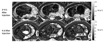 |
42 | Ferumoxytol-Enhanced T2* Mapping of Human Placenta with Fetal Growth Restriction
Ruiming Chen1, Ante Zhu2, Kevin M. Johnson1,3, Ian M. Bird4, Kathleen M. Antony4, J. Igor Iruretagoyena4, Scott B. Reeder1,3,4,5,6, Diego Hernando1,3,7,8, Dinesh M. Shah4, and Oliver Wieben1,3
1Medical Physics, University of Wisconsin - Madison, Madison, WI, United States, 2GE Global Research, Niskayuna, NY, United States, 3Radiology, University of Wisconsin - Madison, Madison, WI, United States, 4Obstetrics & Gynecology, University of Wisconsin - Madison, Madison, WI, United States, 5Medicine, University of Wisconsin - Madison, Madison, WI, United States, 6Emergency Medicine, University of Wisconsin - Madison, Madison, WI, United States, 7Biomedical Engineering, University of Wisconsin - Madison, Madison, WI, United States, 8Electrical and Computer Engineering, University of Wisconsin - Madison, Madison, WI, United States
Fetal growth restriction (FGR) is associated with placental hypoxia, which can be identified by measuring placental T2* value in-vivo. In this pilot study, we acquired T2* maps in 2 subjects in late pregnancy diagnosed with FGR. Mean and histogram of T2* and R2* values in the placenta were analyzed for two timepoints after administration of ferumoxytol reflecting T2* maps dominated by the presence of the iron nanoparticles and a baseline. We observed lower baseline T2* values than previously reported in healthy controls at similar gestation as well as lower baseline T2* values as reported in FGR patients earlier in gestation.
|
||
0818 |
43 | Multiparametric Modeling of Fetal MRI to Compare Lobular and Whole-Placental Segmentations for Fetal Growth Restriction
Janina Schellenberg1, Rosalind Aughwane2, Dimitra Flouri1, Nada Mufti2, Katarzyna Maksym2, Anna L. David2, and Andrew Melbourne1
1Kings College London, London, United Kingdom, 2University College London, London, United Kingdom Babies with fetal growth restriction fail to grow properly in the womb and become hypoxic. They must be delivered prematurely to prevent stillbirth. DECIDE model fitting can determine properties which give information about the placental function and fetal blood saturation. This can be used to identify fetal growth restriction and manage the pregnancy effectively. The aim is to compare DECIDE model fitting results for different mask segmentations. The placenta is segmented into lobules and used as a mask itself. Statistically significant differences of the fetal and placental properties between appropriately developing and growth-restricted pregnancies are determined. |
||
0819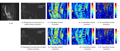 |
44 | Impact of motion correction on in-vivo assessment of Placenta Adhesion Abnormality (PAA) disorders using IVIM method
Mbaimou Auxence Ngremmadji1, Robin Draveny1, Audrey Krisch2, Khalid Ambarki3, Dominik NICKEL4, Charline Bertholdt1,5, Olivier Morel1,5, Marine Beaumont1,2, and Bailiang Chen1,2
1IADI, Inserm U1254, Université de Lorraine, Nancy, France, 2CIC-IT 1433, Inserm, CHRU Nancy, Nancy, France, 3Siemens Healthineers, Nancy, France, 4MR Applications Predevelopment, Siemens Healthcare GmbH, Erlangen, Germany, 5Women's Division, Nancy Regional and University Hospital Center (CHRU), Université de Lorraine, Nancy, France, Nancy, France
Placenta adhesion abnormality (PAA) is the consequence of an excessive invasion of the placenta within the myometrium. Due to its severe maternal pregnancy outcomes, screening of subjects with suspicions of PAA are very important. The utilisation of intra voxel incoherent motion (IVIM) technique may help to improve the diagnosis. But its stability and robustness need to be improved and analysed. Here we aim to evaluate the impact and address the importance of motion correction technique to assess PAA disorders in-vivo using IVIM method in order to establish a robust and stable pipeline for PAA patient screening.
|
||
0820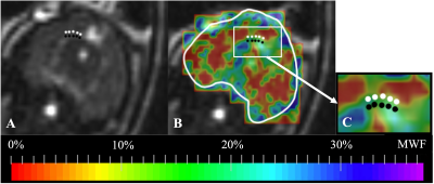 |
45 | Feasibility of Quantifying Myelin Water Fraction in the Fetal Guinea Pig Brain
Simran Sethi1, Lanette J. Friesen-Waldner1, Trevor P. Wade1,2, Ousseynou Sarr3, Brian Sutherland3, Timothy R.H. Regnault3,4,5, and Charles A. McKenzie1,2,5
1Medical Biophysics, Western University, London, ON, Canada, 2Robarts Research Institute, Western University, London, ON, Canada, 3Physiology and Pharmacology, Western University, London, ON, Canada, 4Obstetrics and Gynaecology, Western University, London, ON, Canada, 5Maternal, Fetal & Newborn Health, Children's Health Research Institute, Lawson Research Institution, London, ON, Canada
Myelin water fraction (MWF), a validated marker for myelin lipid, can be used to assess fetal myelination in vivo. Thus, this study aims to demonstrate the feasibility of quantifying fetal MWF. 11 pregnant guinea pigs with 38 fetuses were imaged in a 3T MRI. mcDESPOT was used to quantify MWF in the corpus callosum (CC) of the maternal brain and the CC and fornix (FOR) of the fetal brain. MWF maps were successfully generated in all fetal brains, confirming its feasibility. Maternal MWF results and comparison of fetal CC and FOR MWF results are consistent with published results.
|
||
0821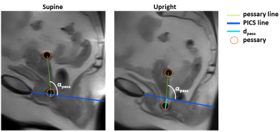 |
46 | Assessment of pessary position using upright MR imaging of patients with pelvic organ prolapse
Lisan M. Morsinkhof1, Anique T.M. Grob2, Lianne Straetemans1, and Frank F.J. Simonis1
1Magnetic Detection & Imaging, University of Twente, Enschede, Netherlands, 2Multi-Modality Medical Imaging, University of Twente, Enschede, Netherlands
Pessary treatment for pelvic organ prolapse (POP) has a success rate of only 63% and its working mechanism remains unclear. This study applies upright MRI to gain insight into pessary. Patients were scanned in supine and upright position. The pessary was located under the inferior pubic bone in 7 out of 9 patients in upright position, opposed to 1 out of 9 in supine position. This indicates that the pubic bone does not (solely) contribute to the supporting mechanism. The use of upright MRI contributes to knowledge about the working mechanism of the pessary.
|
||
0822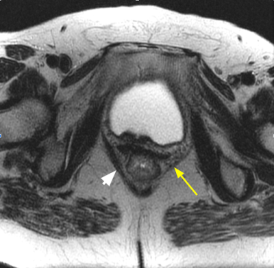 |
47 | Role of puborectalis thickness measurement in the assessment of pelvic floor dysfunction and its correlation with HMO classification.
Anugayathri Jawahar1, Perla Subbaiah 2, and Vipul R Sheth3
1Radiology, Loyola University Medical Center, Maywood, IL, United States, 2Biostatistics, Oakland University, Rochester, MI, United States, 3Radiology Body MRI, Stanford University Medical Center, Palo Alto, CA, United States
Pelvic floor dysfunction is a common problem affecting women over 50 years of life causing significant morbidity. Traditionally, H-line and M-line measurements have been used to assess pelvic organ prolapse. However, puborectalis muscle which plays a crucial role in pelvic floor intactness can be measured to reflect the weakening of the pelvic floor. We evaluated and found puborecatalis measurement can be used as a supplement to H-line and M-line measurements in assessing pelvic organ prolapse.
|
||
The International Society for Magnetic Resonance in Medicine is accredited by the Accreditation Council for Continuing Medical Education to provide continuing medical education for physicians.