Digital Poster
Female Pelvis II: Gynecologic Cancers
Joint Annual Meeting ISMRM-ESMRMB & ISMRT 31st Annual Meeting • 07-12 May 2022 • London, UK

| Computer # | ||||
|---|---|---|---|---|
0915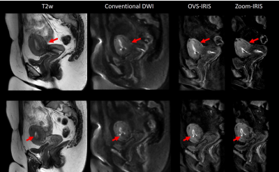 |
35 | Feasibility of high-quality uterus DWI with segmented phase-encoding and reduced FOV Video Not Available
Yajing Zhang1, Zhigang Wu2, Xiuquan Hu3, Guangyu Jiang4, Jing Zhang3, Jiazheng Wang3, and Yan Zhao5
1MR Clinical Science, Philips Healthcare, Suzhou, China, 2Philips Healthcare (China), Shenzhen, China, 3Philips Healthcare (China), Beijing, China, 4MR Clinical Application, Philips Healthcare, Suzhou, China, 5MR R&D, Philips Healthcare, Suzhou, China Diffusion-weighted imaging (DWI) of uterus has been crucial for providing useful information in differentiating pathologies such as uterine fibroids and adenomyosis. In this study, we aim to investigate the feasibility of high quality uterus DWI through a segmented phase-encoding acquisition with reduced FOV. |
||
0916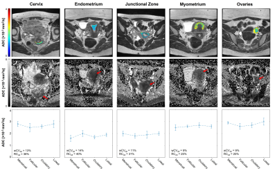 |
36 | Repeatability of Apparent Diffusion Coefficient Estimates of Healthy Male and Female Pelvic Tissues
Lauren K Fang1, Ana E Rodríguez-Soto1, Anders M Dale1,2, Christopher C Conlin1, Tyler M Seibert1,3,4, Michael E Hahn1, and Rebecca A Rakow-Penner1,4
1Radiology, University of California, San Diego, La Jolla, CA, United States, 2Neuroscience, University of California, San Diego, La Jolla, CA, United States, 3Radiation Medicine and Applied Sciences, University of California, San Diego, La Jolla, CA, United States, 4Bioengineering, University of California, San Diego, La Jolla, CA, United States Establishing ADC repeatability is important for clinical translation of quantitative DWI. Pelvic ADC repeatability in healthy men and premenopausal women are not well-defined. Four men were scanned twice and six women were scanned each week of their normal 28-day menstrual cycle using a 3.0T scanner. Prostate, cervix, and uterine tissue ROIs were drawn to calculate median ADC values. Minimum reliably detectable ADC change (RC) was estimated at 16% and 11% within the prostatic peripheral and transition zones, respectively. Female reproductive tissue ADCs and repeatability varied across the menstrual cycle, with the greatest RCs displayed by endometrium (40%) and cervix (36%). |
||
0917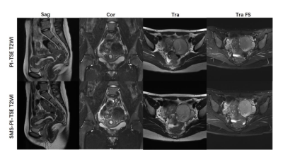 |
37 | Performance Evaluation of high resolution T2-TSE sequence based on simultaneous multi-slice (SMS) in Female Pelvis MRI Video Not Available
Hongchao Wang1, Yueluan Jiang2, Zhuo Wang1, Lei Zhang1, Zhiqing Shao1, Yang Sun1, and Xiaoye Wang3
1The First Hospital of Jilin University, Changchun, China, 2MR Scientific Marketing, Siemens Healthineers, Beijing, China, 3MR Clinical Marketing, Siemens Healthineers, Beijing, China
Simultaneous multi-slice (SMS) acquisition is a different option for accelerating 2D TSE MRI through the excitation of multiple slices simultaneously and is associated with much smaller SNR loss than parallel acceleration. In this study, we applied both parallel imaging and multiple slices simultaneous imaging technique to accelerate T2WI TSE of female pelvis MRI. Comparing to conventional TSE sequence, 25% of time could be reduced with more slices with thinner thickness using SMS acceleration with similar performances at 3T. SMS-PI-TSE is a feasible acceleration technique in Pelvis MRI.
|
||
0918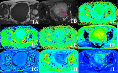 |
38 | Multiple quantitative parameters diagnosis of cellular and degeneration uterine leiomyoma with non-enhancement MRI Video Not Available
Shifeng Tian1 and Ailian Liu1,2
1the First Affiliated Hospital of Dalian Medical University, Dalian, China, 2Dalian Medical Imaging artificial intelligence engineering technology research center, Dalian, China
Results shown that several quantitative parameters of diffusion kurtosis imaging (DKI) and enhanced T2 star weighted angiography (ESWAN) sequence had different between cellular uterine leiomyoma (CUL) and degeneration uterine leiomyoma (DUL). Therefore, it is feasible to use DKI and ESWAN to distinguish CUL and DUL.
|
||
0919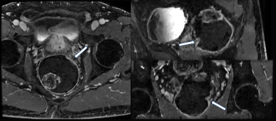 |
39 | Rapid and High Resolution Pelvic MRI Using Deep Learning Reconstruction
Melany B Atkins1, Arnaud Guidon2, Michael Vinski3, Thomas Schrack4, Heidi Harris5, and Ersin Bayram6
1Radiology, Fairfax Radiological Consultants, Arlington, VA, United States, 2GE Healthcare, Boston, MA, United States, 3GE Healthcare, Lynchburg, VA, United States, 4Fairfax Radiological Consultants, Fairfax, VA, United States, 5GE Healthcare, Waukesha, WI, United States, 6GE Healthcare, Houston, TX, United States
MRI plays an important role in pelvic assessment. For instance, it is the modality of choice for rectal cancer staging, gynecologic cancer staging, uterine fibroid evaluation and ovarian tumor characterization. Due to the complex nature of the anatomy and clinical demands of these protocols, high resolution thin slice volumetric scans are desired but low SNR, prolonged scan times and motion artifacts remain problematic. In this work, we deploy a combination of recent technical advances in particular high density flexible coils, compressed sensing, and deep learning reconstruction to tackle these challenges and report our feasibility results.
|
||
0920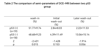 |
40 | The value of semi-quantitative parameters of Dynamic Contrast-Enhanced MRI(DCE-MRI) to predict the expression of P53 in the ovarian cancer
Xulun Lu1, Ye Li1, Qingling Song1, and Ailian Liu1
1First Affiliated Hospital of Dalian Medical University, Dalian, China
P53 is a tumor suppressor gene,TP53-mutated tumors in general have an aggressive phenotype and are characterized by poor differentiation, increased invasiveness, and high metastatic potential[1]。The early enhancement rate reflects the blood perfusion in the early enhancement stage of the lesion. Tumor exhibits a higher early enhancement rate with more abundant the blood supply of the tumor. Our study proved that the wash-in rate of ovarian cancer in the p53 positive group was higher than that in the P53 negative group.
|
||
0921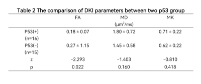 |
41 | A Pilot Evaluation of DKI in Characteristics and Diagnosis of Ovarian Cancer of p53 Status
Shiqi Chen1, Yulin Chen1, Ye Li1, and Ailian Liu1
1the First Affiliated Hospital of Dalian Medical University, Dalian, China
The identification and treatment of ovarian cancer(OC) has always been a problem that plagues us[1]. Mutations in the p53 gene showed significantly up-regulation in human OC tissues and was closely associated with the development of human OC. This work aimed at exploring the value of Diffusion kurtosis imaging (DKI) for characteristics and diagnosis of ovarian cancer of p53 status. The results showed that FA provided a promising performance (AUC =0.742 , sensitivity = 100%, specificity = 43.7%) in evaluating p53 status and OC.
|
||
0922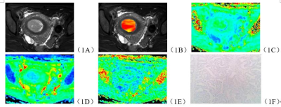 |
42 | Evaluation of microsatellite instability in endometrial carcinoma by amide proton transfer imaging and diffusion kurtosis imaging Video Not Available
Shifeng Tian1, Ailian Liu1,2, Xing Meng1, Lihua Chen1, Nan Wang1, Liangjie Lin3, Geli Hu3, and Jiazheng Wang3
1the First Affiliated Hospital of Dalian Medical University, Dalian, China, 2Dalian Medical Imaging artificial intelligence engineering technology research center, Dalian, China, 3Philips Healthcare, Beijing, China
Microsatellite instability (MSI) is one of the four types of endometrial carcinoma (EC) molecular classification based on The Cancer Genome Atlas (TCGA) [1], which refers to the high mutation load caused by the failure of EC to correct certain errors in small sequence replication due to the abnormal function of DNA mismatch repair system [2]. The purpose of this study is to explore the value of multiple quantitative parameters of APT and DKI sequences in evaluating EC MSI status.
|
||
0923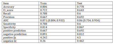 |
43 | Application value of radiomics methods based on DTI sequence FA map for differentiating squamous cell carcinoma from cervix adenocarcinoma Video Not Available
Changjun Ma1, Shifeng Tian2, and Ailian Liu2
1Department of Radiology, the First Affiliated Hospital of Dalian Medical University, Dalian,China, China, 2Department of Radiology, the First Affiliated Hospital of Dalian Medical University, Dalian, China
Through high-throughput quantitative feature extraction, the imaging omics method achieves non-invasive analysis of tumor heterogeneity and improves the accuracy of imaging examination in assisting clinical decision making in tumor screening, diagnosis and prognosis prediction. This study intends to explore the value of imaging omics method based on DTI sequence FA map in differentiating CSCC and CA, and provide imaging basis for the formulation of individualized precision treatment plan for CC patients.
|
||
0924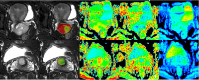 |
44 | The value of APTw combined with DKI for prediction of pelvic lymph node metastasis of cervical cancer Video Not Available
Xing Meng1, Ailian Liu1,2, Shifeng Tian1, Changjun Ma1, Liangjie LIN3, Qingwei Song1, Geli Hu3, and Jiazheng Wang3
1the First Affiliated Hospital of Dalian Medical University, Dalian, China, 2Dalian Medical Imaging artificial intelligence engineering technology research center, Dalian, China, 3MSC Clinical & Technical Solutions, Philips Healthcare, Beijing, China
Cervical cancer is one of the most common malignant cancers in pelvic cavity, and trends in young adults. This study aims to investigate the value of amide proton transfer weighted (APTw) combined with diffusion kurtosis imaging (DKI) for prediction of pelvic lymph node metastasis of cervical cancer, the accurate diagnosis of which is significant for the treatment and prognosis of cervical cancer.
|
||
The International Society for Magnetic Resonance in Medicine is accredited by the Accreditation Council for Continuing Medical Education to provide continuing medical education for physicians.