Digital Poster
Sequences & Sampling I
Joint Annual Meeting ISMRM-ESMRMB & ISMRT 31st Annual Meeting • 07-12 May 2022 • London, UK

| Computer # | ||||
|---|---|---|---|---|
1611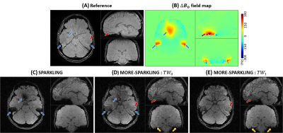 |
66 | MORE-SPARKLING: Non-Cartesian trajectories with Minimized Off-Resonance Effects
Chaithya G R1,2, Guillaume Daval-Frérot1,2,3, Aurélien Massire3, Boris Mailhe4, Mariappan Nadar4, Alexandre Vignaud1, and Philippe Ciuciu1,2
1NeuroSpin, Joliot, CEA, Université Paris-Saclay, F-91191, Gif-sur-Yvette, France, 2Inria, Parietal, Université Paris-Saclay, F-91120, Palaiseau, France, 3Siemens Healthcare SAS, Saint-Denis, 93210, France, 4Siemens Healthineers, Princeton, NJ, United States We augment the recently introduced SPARKLING algorithm and propose an improved mathematical formulation that takes the temporal dependence of the MR signal into account. This prevents the trajectories from sampling similar portions of k-space at different times, thereby reducing distortions and blurring induced by \(B_0\) inhomogenieties. Overall, these trajectories present a smooth distribution over time in k-space and Minimized Off-Resonance Effects (MORE-SPARKLING), verified both retrospectively and prospectively with scans performed in vivo at 3T on a healthy volunteer. |
||
1612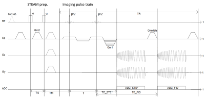 |
67 | Spiral 3DREAM sequence for fast whole-brain B1 Mapping
Marten Veldmann1, Svenja Niesen1, Philipp Ehses1, and Tony Stöcker1,2
1Deutsches Zentrum für Neurodegenerative Erkrankungen e.V. (DZNE), Bonn, Germany, 2Department of Physics & Astronomy, University of Bonn, Bonn, Germany
The 3DREAM sequence provides a fast technique for whole-brain B1 mapping, but suffers from different blurring levels in FID and STE* images. We developed a new variant of the 3DREAM sequence with a spiral readout to mitigate this problem. The spiral readout shortens the echo train duration and reduces the number of excitations after the STEAM preparation significantly. This leads to similar blurring levels in FID and STE* images and increases the effective resolution of flip angle maps. As a result, correction strategies for different blurring levels are no longer necessary.
|
||
1613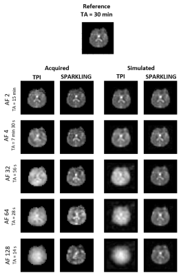 |
68 | Evaluation of 3D SPARKLING readout for Sodium UTE MRI at ultra-high magnetic field
Renata Porciuncula Baptista1, Alexandre Vignaud1, Chaithya G R1,2, Guillaume Daval-Frérot1,2, Franck Mauconduit1, Mathieu Naudin3, Marc Lapert4, Remy Guillevin3, Philippe Ciuciu1,2, Cécile Rabrait-Lerman1, and Fawzi Boumezbeur1
1NeuroSpin, Joliot, CEA, CNRS, University Paris-Saclay, Gif-sur-Yvette, France, 2Inria, Parietal, Université Paris-Saclay, Palaiseau, France, 3University Hospital Centre Poitiers, DACTIM-MIS, Poitiers, France, 4Siemens Healthcare SAS, Saint-Denis, France
Quantitative 23Na MRI provides useful information about brain tissue homeostasis. Most sequences use deterministic non-Cartesian 3D trajectories such as TPI. However, stochastic strategies such as SPARKLING could improve the coverage of k-space. This study evaluates the advantages of SPARKLING versus TPI. From in vivo datasets at 7T, we determined that undersampled SPARKLING acquisitions outperform TPI for (8 mm)3 resolution with a birdcage coil or for (4 mm)3 with a 32-channel coil. Through extrapolation of these results, we predict that at 11.7T/32-channel, 23Na MRI data could be acquired in 90s at (3 mm)3, which could be interesting for sodium fMRI imaging.
|
||
1614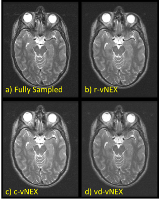 |
69 | SNR Enhancement of MR Images with Variable Compress Sensed Excitation: vNEX
Harsh Kumar Agarwal1, Shaik Ahmed1, Rajagopalan Sundaresan1, Sudhanya Chatterjee1, Sajith Rajamani1, Ashok Kumar P Reddy1, Bhairav Mehta1, Dattesh Shanbhag1, and Ramesh Venkatesan1
1GE Healthcare, Bangalore, India
MR imaging is signal starve leading to long acquisition time. Multiple averaging/excitation is most common way to boost the SNR. However, it is associated with proportionately long scan time. This abstract presents Variable Compress Sensed Excitation (vNEX) image acquisition and image reconstruction technique which subsample each signal average by compressed sensing fast MRI technique and robust image reconstruction is done to reconstruct MRI images for individual signal averages. The proposed technique is demonstrated for T2w brain MRI where SNR equivalence of 336sec scan time is shown in a 200sec vNEX MRI. Three sampling patterns were proposed and statistically compared.
|
||
1615 |
70 | MRI with Sub-Millisecond Temporal Resolution: SPIRE Imaging of a High-speed Motion Phantom
Fischer Johannes1 and Michael Bock1
1Dept. of Radiology, Medical Physics, Medical Center University of Freiburg, Faculty of Medicine, Uni, Freiburg, Germany
Imaging sequences with sub-millisecond temporal resolution based on single point imaging (SPI) are very time inefficient. We designed and imaged a motion phantom capable of velocities up to 5.5m/s. A synchronization signal is acquired using a photoelectric barrier and combined with a sequence trigger signal. Reconstructed images show clear delineation of small structures with a spatial resolution of $$$\Delta x$$$=0.67mm and a temporal resolution of $$$\delta t$$$=246μs.
|
||
1616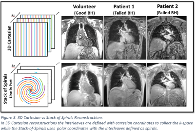 |
71 | Spiral UTE MRI of the Lung: An Investigation on Motion Sensitivity
Valentin Fauveau1, Adam Jacobi2, Adam Bernheim2, Michael Chung2, Yang Yang1,2, Thomas Benkert3, Zahi Fayad1,2, and Li Feng1,2
1BioMedical Engineering and Imaging Institute, Icahn School of Medicine at Mount Sinai, New York, NY, United States, 2Department of Radiology, Icahn School of Medicine at Mount Sinai, New York, NY, United States, 3MR Application Predevelopment, Siemens Healthcare GmbH, Erlangen, Germany
Spiral UTE is a relatively new MRI technique that combines ultra-short echo time acquisition with a stack-of-spirals trajectory for imaging the lung. In this work, we aimed to analyze the motion sensitivity of different reordering schemes and breath holding positions in spiral UTE MRI to determine the optimized protocol for imaging the lungs. A blind assessment was performed by three experienced chest radiologists and the results were analyzed with statistical tests. The Spiral UTE with line-in-partition reordering performed during the inspiration phase was considered the best protocol in our study.
|
||
1617 |
72 | SPEN, RESOLVE and Spin-Echo EPI at 7T: A single-Tx human brain scan comparison
Martins Otikovs1 and Lucio Frydman1,2
1Department of Chemical and Biological Physics, Weizmann Institute of Science, Rehovot, Israel, 2Azrieli National Center for Brain Imaging, Weizmann Institute of Science, Rehovot, Israel
Spatiotemporal encoding (SPEN) is an ultrafast imaging technique, that can serve as useful alternative to single shot and to read-out segmented (RESOLVE) spin-echo (SE) EPI methods, when trying to overcome image distortions due to magnetic field (B0) inhomogeneities. SPEN’s reliance on adiabatic sweeps could also make it an attractive candidate for imaging at high fields, and overcoming B1+-related inhomogeneities. Herein we present phantom and in-vivo results acquired on a 7T human imaging system for single and multi-shot SPEN acquisitions, corroborating its robustness vs comparable single and multi-shot SE-EPI and RESOLVE acquisitions implemented on a single-Tx configuration.
|
||
1618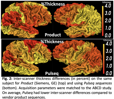 |
73 | A vendor-neutral 3D MP-RAGE implementation in Pulseq for reduced inter-site variability
Yogesh Rathi1, Jon-Fredrik Nielsen2, Maxim Zaitsev3, Lipeng Ning1, Jr-Yuan Chiou4, Tae Hyung Kim5, William Grissom6, Borjan Gagoski7, and Berkin Bilgic5
1Psychiatry, Brigham and Women's Hospital, Harvard Medical School, Boston, MA, United States, 2University of Michigan, Ann Arbor, MI, United States, 3High Field MR Center, Center for Medical Physics and Biomedical Engineering, Medical University of Vienna, Vienna, Austria, 4Brigham and Women's Hospital, Boston, MA, United States, 5Athinoula A. Martinos Center for Biomedical Imaging, Harvard Medical School, Boston, MA, United States, 6Vanderbilt University, Nashville, TN, United States, 7Fetal-Neonatal Neuroimaging and Developmental Science Center, Boston Children’s Hospital, Harvard Medical School, Boston, MA, United States
Inter-scanner variability can reduce the statistical power of neuroimaging studies due to the increased variance in the data. While existing efforts employ consistent high level parameters to acquire data across scanners, we hypothesize that differences due to sequence implementation and reconstruction algorithms contribute substantially to variability in multi-center settings. We propose a unified vendor-neutral MP-RAGE protocol based on the Pulseq sequence development platform that can be used consistently across sites and vendors for reducing inter-scanner variability. Preliminary results show increased consistency of cortical thickness across sites using Pulseq protocol compared to vendor-provided sequences.
|
||
1619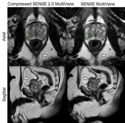 |
74 | Fast and motion robust 2D T2 TSE Propeller acquisition of the prostate with Compressed SENSE: comparison with conventional SENSE acquisition
Leon Bischoff1, Christoph Katemann2, Oliver Weber2, Alexander Isaak1, Dmitrij Kravchenko1, Narine Mesropyan1, Christoph Endler1, Thomas Vollbrecht1, Claus Christian Pieper1, Ulrike Attenberger1, and Julian Luetkens1
1Department of Diagnostic and Interventional Radiology, University Hospital Bonn, Bonn, Germany, 2Philips GmbH Market DACH, Hamburg, Germany
Multiparametric MRI (mpMRI) of the prostate can detect clinically significant prostate cancer in men. To accelerate the time-consuming acquisition process, we integrated a new Compressed SENSE (CS) method for T2-weighted sequences with propeller acquisition and compared it qualitatively and quantitatively to conventional SENSE accelerated T2-weighted propeller sequences. We could demonstrate that while the new CS acquisition method has less artifacts, better image sharpness and higher apparent signal-to-noise ratio (aSNR) and contrast-to-noise ratio (CNR), it reduced the acquisition time by 24%. These findings indicate a superiority over the conventional T2-sequence and could improve the diagnostic workup of patients with prostate cancer.
|
||
1620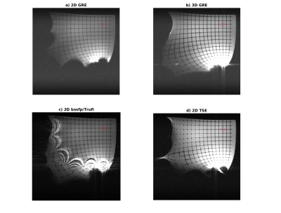 |
75 | Imaging Off-Center: How far can we move outside the central imaging region
Sebastian Littin1, Patrick Hucker1, Maximilian Frederik Russe2, Feng Jia1, Philipp Amrein1, and Maxim Zaitsev1,3
1Department of Radiology, Medical Physics, University Freiburg, Faculty of Medicin, Freiburg, Germany, 2Department of Radiology, University Freiburg, Faculty of Medicin, Freiburg, Germany, 3Center for Medical Physics and Biomedical Engineering, High Field MR Center, Medical University of Vienna, Vienna, Austria Imaging outside the predefined and optimized FoV may enhance accessibility for interventional MRI procedures. Here we present different measures to assess the encoding capability of gradient systems in off-center regions. Different clinically establishes sequences are compared at 0.55T and 1.5T regarding their imaging capabilities beyond the predefined target regions. |
||
1621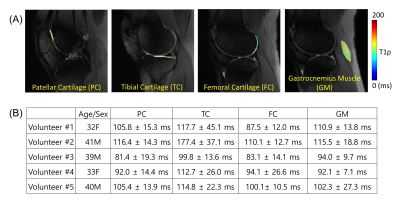 |
76 | Feasibility of 3D Adiabatic T1ρ-prepared Fast Spin Echo (3D Adiab-T1ρ-FSE) Imaging
Hyungseok Jang1, Yajun Ma1, Dina Moazamian1, Michael Carl2, Saeed Jerban1, Alecio F Lombardi1, Christine B Chung1,3, Eric Y Chang1,3, and Jiang Du1
1Radiology, University of California San Diego, San Diego, CA, United States, 2GE Healthcare, San Diego, CA, United States, 3Radiology Service, Veterans Affairs San Diego Healthcare System, San Diego, CA, United States
T1ρ has been investigated as a quantitative biomarker sensitive to changes in macromolecules such as proteoglycan and collagen in musculoskeletal systems. More recently, adiabatic T1ρ (Adiab-T1ρ) has emerged as an alternative to conventional continuous wave T1ρ to reduce the magic angle effect, which is a major confounding factor in accurate T1ρ estimation. In this study, we investigated a new pulse sequence combining adiabatic T1ρ preparation and efficient 3D fast spin echo (FSE) for more robust adiab-T1ρ mapping in the human knee. The efficacies of RF cycling and magnetization reset were also demonstrated.
|
||
The International Society for Magnetic Resonance in Medicine is accredited by the Accreditation Council for Continuing Medical Education to provide continuing medical education for physicians.