Digital Poster
Emerging Techniques in Data Acquisition & Analysis I
Joint Annual Meeting ISMRM-ESMRMB & ISMRT 31st Annual Meeting • 07-12 May 2022 • London, UK

Digital Poster
Emerging Techniques in Data Acquisition & Analysis I
| Computer # | ||||
|---|---|---|---|---|
0866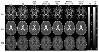 |
91 | Optimized denoising and removal of partial-Fourier induced Gibbs ringing improves accuracy and robustness of DTI and DKI parameters
Jenny Chen1, Benjamin Ades-aron1, Hong-Hsi Lee2, Durga Kullakanda1, Saurabh Maithani1, Dmitry S. Novikov1, Jelle Veraart1, and Els Fieremans1
1Department of Radiology, NYU School of Medicine, New York, NY, United States, 2Athinoula A. Martinos Center for Biomedical Imaging, Massachusetts General Hospital, Boston, MA, United States
Diffusion MRI (dMRI) is affected by noise and by artifacts such as Gibbs ringing and distortions. Using software phantoms as ground-truth, this study compares diffusion tensor imaging (DTI) and diffusional kurtosis imaging (DKI) parameter estimates to assess accuracy of various denoising and Gibbs removal methods, two key components of dMRI pipelines. An optimized Diffusion parameter EStImation with Gibbs and NoisE Removal (DESIGNER) pipeline is proposed, with non-local patch MP-PCA denoising and Removal of Partial-fourier induced Gibbs Ringing (RPG), that yields more accurate metrics, fewer outlier voxels in phantoms and more robust DTI/DKI maps in patient data.
|
||
0867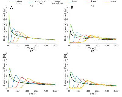 |
92 | Comparison of image and extracorporeal derived arterial input functions for DCE-MRI in mice using an multimodal cross-validation approach
Florian Gierse1, Juela Cufe1, Bastian Maus2, Katharina Kronenberg3, Michael Claesener4, Paulina Dorten1, Klaus Schäfers1, Sven Hermann1, Uwe Karst3, Cornelius Faber2, Michael Schäfers4, Florian Büther4, and Philipp Backhaus4
1European Institute for Molecular Imaging, University of Münster, Münster, Germany, 2Translational Research Imaging Center, University of Münster, Münster, Germany, 3Institute of Inorganic and Analytical Chemistry, University of Münster, Münster, Germany, 4Department of Nuclear Medicine, University Hospital Münster, Münster, Germany Quantitative precise measurements of the dynamic arterial blood concentration (AIF) are challenging in perfusion MRI. As solution an extracorporeal circulation approach for the AIF determination with two different vascular access ways is presented and cross validated with injection of radioactive contrast agent derivatives. Independent of the vascular access way, extracorporeal derived AIFs point towards a high quantitative precision, although dispersion must be considered. However, deconvolution methods based on single gamma variate functions end up in plausible AIFs. The use of novel rapid imaging techniques in connection with extracorporeal AIFs indicates high potential for precise quantitative dual PET/MRI Pharmacokinetic-Modeling in mice. |
||
| 0868 | 93 | Mapping Rodent Brain Mechanical Properties In Vivo with Magnetic Resonance Elastography and Nonlinear Inversion
L. Tyler Williams1, Katrina A. Milbocker2, Seth R. Sullivan1, Ian F. Smith2, Eric Brengel2, Gillian LeBlanc2, Anna Y. Klintsova2, Matthew D. J. McGarry3, and Curtis L. Johnson1
1Biomedical Engineering, University of Delaware, Newark, DE, United States, 2Psychological & Brain Sciences, University of Delaware, Newark, DE, United States, 3Thayer School of Engineering, Dartmouth College, Hanover, NH, United States
Developing preclinical MRE techniques that are comparable to human MRE is important for translational studies. This pilot study investigates the efficacy of the nonlinear inversion algorithm on rat brain MRE data. Whole-brain MRE scans were performed on female, Long-Evans rats using a custom MRE-EPI sequence and piezoelectric actuator with a resolution of 0.25 mm isotropic and with 600 Hz vibration. NLI parameters were adjusted for brain size and frequency. The resulting shear stiffness and damping ratio maps exhibited strong contrast between different anatomical regions. These results validate the use of NLI in preclinical MRE settings.
|
||
0869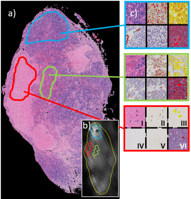 |
94 | VERDICT MRI for tumor tissue characterization – separating viable tumor from necrosis
Mikael Montelius1, Lukas Lundholm1, Oscar Jalnefjord1,2, Eva Forssell-Aronsson1,2, and Maria Ljungberg1,2
1Medical radiation sciences, Institute of Clinical Sciences, University of Gothenburg, Gothenburg, Sweden, 2Medical Physics and biomedical engineering, Sahlgrenska University Hospital, Gothenburg, Sweden
The biological meaning of MRI-based biomarkers of cancer can be studied using spatially matched histologically stained tumor sections. However, correlations are often weak or contradictory, possibly due to confounding necrosis that models fail to describe. In this work, we demonstrate the potential of using VERDICT MRI model parameters to separate necrosis from viable tumor tissue non-invasively. Radiation treated tumors demonstrating large necrotic and viable regions are imaged, excised and stained using immunohistochemical methods. Correlations between spatially matched VERDICT and immunohistochemical maps reveal that VERDICT clearly separates necrosis from viable tumor.
|
||
 Back to Meeting Home
 Back to the Program-at-a-Glance
The International Society for Magnetic Resonance in Medicine is accredited by the Accreditation Council for Continuing Medical Education to provide continuing medical education for physicians.
 Back to Meeting Home
 Back to the Program-at-a-Glance
 View the Presentation