Digital Poster
Spectroscopy II
Joint Annual Meeting ISMRM-ESMRMB & ISMRT 31st Annual Meeting • 07-12 May 2022 • London, UK

| Computer # | ||||
|---|---|---|---|---|
2610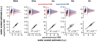 |
51 | Linear-combination modeling of short-TE PRESS data using parameterized and experimentally derived macromolecule basis functions
Helge Jörn Zöllner1,2, Tao Gong3,4, Steve C. N. Hui1,2, Yulu Song1,2, Weibo Chen5, Guangbin Wang3,4, Georg Oeltzschner1,2, and Richard A. E. Edden1,2
1Russell H. Morgan Department of Radiology and Radiological Science, The Johns Hopkins University School of Medicine, Baltimore, MD, United States, 2F. M. Kirby Research Center for Functional Brain Imaging, Kennedy Krieger Institute, Baltimore, MD, United States, 3Departments of Radiology, Shandong Provincial Hospital Affiliated to Shandong First Medical University, Jinan, Shandong, China, 4Departments of Radiology, Shandong Provincial Hospital, Cheeloo College of Medicine, Jinan, Shandong, China, 5Philips Healthcare, Shanghai, China
Recent expert consensus emphasizes the use of measured MM spectra for reliable linear-combination modeling (LCM) of short-TE MRS data. Here we compare the metabolite estimates from short-TE PRESS data modeled with a parameterized and measured MM basis functions in a large dataset.
|
||
2611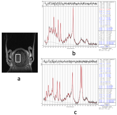 |
52 | Reliable Quantification of Phosphocreatine and total Creatine during Deep Hypothermic Circulatory Arrest in Neonatal Pig Brain using LCModel
Meng Gu1, Ralph Hurd1, and Daniel Spielman1
1Radiology, Stanford University, Stanford, CA, United States
Reliable in-vivo measurement of phosphocreatine using 1H MRS offers valuable information for understanding brain energy metabolism. At 3T, separate quantification of phosphocreatine and creatine has been challenging using LCModel. This problem exacerbates in studies of deep hypothermic circulatory arrest (DHCA), a technique used in many cardiac surgeries, due to temperature-dependent chemical shifts. Furthermore, total creatine quantification is hampered using a 37°C-only basis set as it fails to account for the increased polarization at lower temperatures. By using a semi-laser sequence with temperature dependent LCModel basis sets, reliable quantification of phosphocreatine and total creatine was achieved for the DHCA studies.
|
||
2612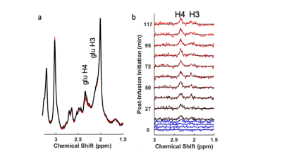 |
53 | 1H spectroscopy of glutamate kinetics with 13C-glucose infusion in rodents at 7T
Christopher D. Kroenke1, Natalie M. Zahr2,3, Evan Kittle3, and Adolf Pfefferbaum2,3
1Oregon Health & Science University, Portland, OR, United States, 2Stanford University, Palo Alto, CA, United States, 3SRI International, Palo Alto, CA, United States
This work implements a method for measuring cerebral kinetics of glucose metabolism using 1H magnetic resonance spectroscopy (MRS) in rats using a small-animal 7T MRI system. The method has been demonstrated to be readily applied to nonhuman primates and human subjects using clinical MRI systems. The extension to small animals requires intravenous administration of small quantities of 13C-labeled glucose, and adaptation of data analysis procedures to data acquired at high magnetic field strength. We demonstrate robust quantification of 13C enrichment within the glutamate at the C4 position. This methodology will facilitate future translational research involving preclinical models of human disease.
|
||
2613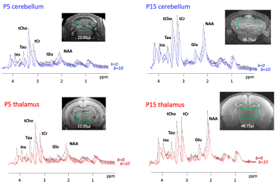 |
54 | Monitoring the complexification of cerebellar and thalamic cells during early development with diffusion-weighted MR spectroscopy
Lily Qiu1, Marco Palombo2,3, Jason Lerch1,4,5, and Clémence Ligneul1
1Wellcome Centre for Integrative Neuroimaging, FMRIB, Nuffield Department of Clinical Neurosciences, University of Oxford, Oxford, United Kingdom, 2Cardiff University Brain Research Imaging Centre (CUBRIC), School of Psychology, Cardiff University, Cardiff, United Kingdom, 3School of Computer Science and Informatics, Cardiff University, Cardiff, United Kingdom, 4Mouse Imaging Centre, The Hospital for Sick Children, Toronto, ON, Canada, 5Department of Medical Biophysics, University of Toronto, Toronto, ON, Canada
Monitoring non-invasively the development of brain cells in the neonate and infant brain could be of interest for neurodevelopmental disorders. In this work, we assess whether diffusion-weighted MR spectroscopy (DWMRS) has the potential to follow cerebellar and thalamic cells development in the healthy rat brain from P5 to P30. Results show that DWMRS is sensitive to microstructural changes in the developing brain. The modeling of the data shows a cell growth and complexification (from P15 to P30), and underlies the need of different modeling hypotheses at earlier time points.
|
||
2614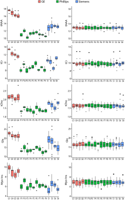 |
55 | Harmonisation of multi-site MRS data with ComBat
Tiffany Bell1,2,3, Kate J Godfrey1,2,3, Ashley L Ware2,3,4,5, Keith Owen Yeates2,3,4, and Ashley D Harris1,2,3
1Department of Radiology, University of Calgary, Calgary, AB, Canada, 2Hotchkiss Brain Institute, University of Calgary, Calgary, AB, Canada, 3Alberta Children's Hospital Research Institute, University of Calgary, Calgary, AB, Canada, 4Department of Psychology, University of Calgary, Calgary, AB, Canada, 5Department of Neurology, University of Utah, Salt Lake City, UT, United States
Multisite magnetic resonance spectroscopy (MRS) studies are becoming increasingly more common, however data collection across multiple sites introduces non-biological variability that can mask true biological effects. Using PRESS and MEGA-PRESS data obtained from the BIG GABA repository and linear modelling, we show that (1) scanner vendor and data collection site is significantly associated with metabolite levels and (2) data harmonisation using ComBat successfully removes non-biological variance to reveal biological effects of interest. We therefore recommend ComBat as an approach to harmonise multi-site MRS data.
|
||
2615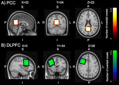 |
56 | Detection of GABA dynamics with edited functional MR spectroscopy at 3 Tesla: a comparison of modelling strategies
Laura Beghini1,2, Francesca Saviola2, Stefano Tambalo2, and Jorge Jovicich2
1Department of Physics, Norwegian University of Science and Technology, Trondheim, Norway, Trondheim, Norway, 2CIMeC, Center for Mind/Brain Sciences, University of Trento, Rovereto (Trento), Italy, Trento, Italy
There is increased interest in measuring brain GABA dynamics with MR spectroscopy, yet there is no consensus about robust and fast ways to do this. In this study, we evaluated how averaging affects GABA estimates when using different modelling strategies (Gannet, Osprey). Our results show that 60 MEGA-PRESS averages give valid concentration estimates, consistent with literature reports. Moreover, Gannet gives a GABA+/Cr measurement which should be more sensitive to concentration variations across regions and give lower temporal variability at rest relative to the GABA+/tCr measure in Osprey.
|
||
2616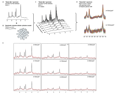 |
57 | Differences in diffusion-weighted MRS processing and fitting pipelines, and their effect on tissue modeling: Results from a workshop challenge.
Chloé Najac1, André Döring2, William Clarke3, Guglielmo Genovese4, Nathalie Just5, Roland Kreis6,7, Henrik Lundell5, Jessie Mosso8,9,10, Eloïse Mougel11, Georg Oeltzschner12,13, Marco Palombo2,14, and Clémence Ligneul3
1C.J. Gorter Center for High-Field MRI, Department of Radiology, Leiden University Medical Center, Leiden, Netherlands, 2Cardiff University Brain Research Imaging Centre (CUBRIC), School of Psychology, Cardiff University, Cardiff, United Kingdom, 3Wellcome Centre for Integrative Neuroimaging, FMRIB, Nuffield Department of Clinical Neurosciences, University of Oxford, Oxford, United Kingdom, 4Center for Magnetic Resonance Research and Department of Radiology, University of Minnesota, Minneapolis, MN, United States, 5Danish Research Centre for Magnetic Resonance, Centre for Functional and Diagnostic Imaging and Research, Copenhagen University Hospital Amager and Hvidovre, Copenhagen, Denmark, 6Magnetic Resonance Methodology, Institute for Diagnostic and Interventional Neuroradiology, University Bern, Bern, Switzerland, 7Translational Imaging Center, sitem-insel, Bern, Switzerland, 8CIBM Center for Biomedical Imaging, Lausanne, Switzerland, 9Animal Imaging and Technology, EPFL, Lausanne, Switzerland, 10LIFMET, EPFL, Lausanne, Switzerland, 11Commissariat à l’Energie Atomique et aux Energies Alternatives (CEA), Centre National de la Recherche Scientifique (CNRS), Molecular Imaging Research Center (MIRCen), Laboratoire des Maladies Neurodégénératives, Université Paris-Saclay, Fontenay-aux-Roses, France, 12Russell H. Morgan Department of Radiology and Radiological Science, The Johns Hopkins University School of Medicine, Baltimore, MD, United States, 13F. M. Kirby Research Center for Functional Brain Imaging, Kennedy Krieger Institute, Baltimore, MD, United States, 14School of Computer Science and Informatics, Cardiff University, Cardiff, United Kingdom
A processing and fitting challenge was initiated for the “Best practices & Tools for Diffusion MR Spectroscopy” workshop held at the Lorentz Center (Leiden, NL) in September 2021. Our goal was to assess variabilities in processing and fitting pipelines and possibly identify the most robust steps to analyze diffusion-weighted MRS data. A “brain-like” dataset of diffusion-weighted spectra was simulated and participants were asked to analyze the data with their routinely used pipelines. Results showed that variabilities between participants (n=8) particularly increased at higher b-values and for J-coupled metabolites (Ins, Glu) as a result of lower SNR and higher CRLB values.
|
||
2617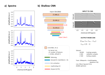 |
58 | Uncertainties and bias in quantification by deep learning in magnetic resonance spectroscopy
Rudy Rizzo1,2, Martyna Dziadosz1,2, Sreenath Pruthviraj Kyathanahally3, and Roland Kreis1,2
1Magnetic Resonance Methodology, Institute of Diagnostic and Interventional Neuroradiology, University of Bern, Bern, Switzerland, 2Translational Imaging Center, sitem-insel, Bern, Switzerland, 3Department Systems Analysis, Integrated Assessment and Modelling, Data Science for Environmental Research group, Dübendorf, Switzerland
Deep Learning has introduced the possibility to speed up quantitation in Magnetic Resonance Spectroscopy. However, questions arise about how to access and relate to prediction uncertainties. Distributions of predictions and Monte-Carlo dropout are here used to investigate data and model related uncertainties, exploiting ground truth knowledge (in-silico set up). It is confirmed that DL is a dataset-biased technique, showing higher uncertainties toward the edges of its training set. Surprisingly, metabolites present in high concentrations suffer from comparable high uncertainties as when present in low concentrations. Evaluating and respecting fitting uncertainties is equally crucial for DL and traditional approaches.
|
||
2618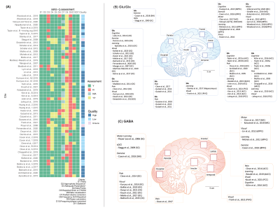 |
59 | A meta-analysis of GABA and Glutamate response in functional magnetic resonance spectroscopy
Duanghathai Pasanta1,2, Georg Oeltzschner3, Talitha Ford4, David J. Lythgoe5, and Nicolaas A. Puts1,6
1Department of Forensic and Neurodevelopmental Sciences, Sackler Institute for Translational Neurodevelopment, Institute of Psychiatry, Psychology, and Neuroscience, King's College London, London, United Kingdom, 2Department of Radiologic Technology, Faculty of Associated Medical Sciences, Chiang Mai University, Chiang Mai, Thailand, 3Russell H. Morgan Department of Radiology and Radiological Science, Johns Hopkins University School of Medicine, Baltimore, MD, United States, 4Cognitive Neuroscience Unit, School of Psychology, Deakin University, Geelong, Australia, 5Department of Neuroimaging, King’s College London, London, United Kingdom, 6MRC Centre for Neurodevelopmental Disorders, King's College London, London, United Kingdom
Functional magnetic resonance spectroscopy (fMRS) can be used to investigate the neurometabolic responses to external stimuli in-vivo, but findings to date are inconsistent. We performed a systematic review and meta-analysis on 49 human fMRS studies on Glutamate, Glx (Glutamate + Glutamine) and GABA. Small to moderate effect sizes of 0.29-0.47 (p < 0.05) were observed for Glu/Glx regardless of stimulus domain but not for GABA. Results suggest Glu/Glx and GABA responses vary with time course and are unique to stimulus domain or task. This analysis will inform study design in future work.
|
||
2619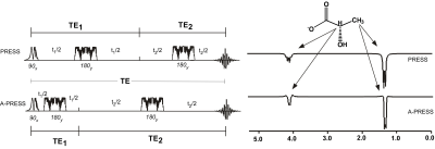 |
60 | The combined use of MEGA- editing and asymmetric-PRESS methods improve the precision of cerebral lactate detection in healthy adults
Lauriane Jugé1,2, Iain Ball3, and Caroline D Rae1,2
1Neuroscience Research Australia, Sydney, Australia, 2School of Medical Sciences, University of New South Wales, Sydney, Australia, 3Philips Australia & New Zealand, North Ryde, Australia
Here, we combined two 1H-MRS methods that have shown utility for detecting lactate in the brain, MEGA- editing and use of an asymmetric-PRESS and compared this approach (MEGA-APRESS) with MEGA-PRESS and A-PRESS in phantoms and eight individuals. MEGA-APRESS showed improved precision compared to MEGA-PRESS. Both editing approaches were greatly superior to A-PRESS alone.
|
||
2620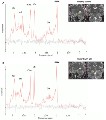 |
61 | Investigation of metabolic changes in the hippocampus following spinal cord injury applying a metabolite-cycling semi-LASER technique
Sandra Zimmermann1, Dario Pfyffer1, Roland Kreis2,3, Kadir Simsek2,3, Patrick Freund1,4, and Maryam Seif1,4
1Spinal Cord Injury Center, Balgrist University Hospital, Zurich, Switzerland, 2Magnetic Resonance Methodology, Institute of Diagnostic and Interventional Neuroradiology, University of Bern, Bern, Switzerland, 3Translational Imaging Center, sitem-insel, Bern, Switzerland, 4Neurophysics Department, Max Plank Institute, Leipzig, Germany
This work uses magnetic resonance spectroscopy data obtained in the hippocampus of 28 patients with traumatic spinal cord injury (SCI) to investigate SCI-induced metabolic changes. Structural magnetic resonance imaging was performed for hippocampal volumetric assessment. Eighteen healthy controls underwent the same imaging protocol. Study participants underwent a functional assessment by testing the visuospatial and verbal memory performance to check for cognitive impairments. In SCI patients without cognitive impairments, hippocampal metabolites did not differ from healthy controls. This study does not support evidence of degeneration and inflammation in the hippocampus of animal models of SCI that show impaired spatial memory performance.
|
||
2621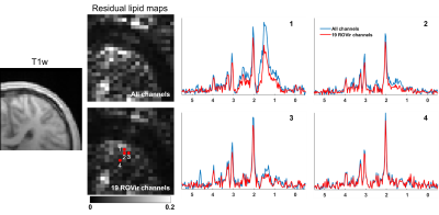 |
62 | Improving Lipid Suppression for 1H-MRSI Using Region-Optimized Virtual Coils
Fan Lam1,2,3, Yahang Li1,2, Yibo Zhao2,4, and Justin Haldar5
1Department of Bioengineering, University of Illinois at Urbana-Champaign, Urbana, IL, United States, 2Beckman Institute for Advanced Science and Technology, University of Illinois at Urbana-Champaign, Urbana, IL, United States, 3Cancer Center at Illinois, University of Illinois at Urbana-Champaign, Urbana, IL, United States, 4Department of Electrical and Computer Engineering, University of Illinois at Urbana-Champaign, Urbana, IL, United States, 5Ming Hsieh Department of Electrical and Computer Engineering, University of Southern California, Los Angeles, CA, United States
One major challenge for 1H-MRSI with localized excitation is minimizing interference from undesired regions, particularly the subcutaneous lipids. In this work, we exploit the complementariness of a new technique called region-optimized virtual coils (ROVir) that is capable of generating a set of virtual channels sensitized to specific regions and spatiospectral processing for lipid removal. Using brain 1H-MRSI data, we demonstrated improved lipid removal by combining ROVir and a state-of-the-art union-of-subspaces method. We expect this integration to be useful for MRSI applications where VOIs are often lateralized (e.g., stroke and tumor) and/or cerebral lipids are of interest.
|
||
The International Society for Magnetic Resonance in Medicine is accredited by the Accreditation Council for Continuing Medical Education to provide continuing medical education for physicians.