Digital Poster
Interventional MRI
Joint Annual Meeting ISMRM-ESMRMB & ISMRT 31st Annual Meeting • 07-12 May 2022 • London, UK

| Computer # | ||||
|---|---|---|---|---|
1085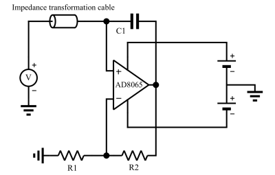 |
92 | A currentless remote decoupling circuit for receive-only endoscopic MRI coils using negative impedance converters
Sina Marhabaie1, Georges Willoquet1, Rose-Marie Dubuisson1, Bruno Quesson2,3,4, and Marie Poirier-Quinot1
1Université Paris-Saclay, BioMaps, Orsay, France, 2IHU L’Institut de RYthmologie et de Modélisation Cardiaque (LIRYC), Electrophysiology and Heart Modeling Institute, Fondation Bordeaux Université, 33600 Pessac, Bordeaux, France, 3Centre de recherche Cardio-Thoracique de Bordeaux, U1045, Université de Bordeaux, 33000, Bordeaux, France, 4INSERM, Centre de Recherche Cardio-Thoracique de Bordeaux, U1045, Université de Bordeaux, 33000, Bordeaux, France Receive-only coils must be decoupled from the transmit coil during excitation. For conventional coils, decoupling is achieved using a resonant trap, which is switched during B1 transmit. However, in situations with very limited space like intravascular coils, this method remains problematic. We aimed to address efficient remote decoupling of a receive-only coil. We implemented an alternative approach by adding a “negative resistance” to the trap. This negative resistance is tailored in such a way that cancels out the positive resistance of the blocking trap, hence augmenting the quality factor of the trap and thus the decoupling efficiency. |
||
1086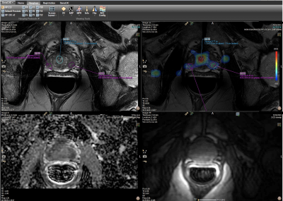 |
93 | New Endo Coil and Novel Methods for Improving mpMR-TRUS Guided Fusion Prostate Biopsies with Hyperpolarized C-13 Pyruvate Molecular Imaging
Daniel Tewelde Gebrezgiabbhier1,2, Hsin-Yu Chen3, Robert Bok3, Lucas Carvajal3, Matthew Cooperberg2, Hao Nguyen2, Katsuto Shinohara2, Kimberly Okamoto3, Mary Frost3, Zhen Wang3, Michael Ohliger3, Jeremy Gordon3, Peder Larson3, Rahul Aggarwal2, and Daniel Vigneron3
1Bioengineering, University of California, San Francisco, CA, United States, 2School of Medicine, University of California, San Francisco, CA, United States, 3Radiology and Biomedical Imaging, University of California, San Francisco, CA, United States
A new 13C/1H endorectal coil was designed, 3D printed, and manufactured with optimal dimensions to improve the comfort and tolerability for patients and provide higher SNR over the original endorectal coil used for over a decade. After bench and phantom tests, the new endorectal coil was applied in patient studies for mpMRI-TRUS guided fusion prostate biopsies with hyperpolarized C-13 pyruvate molecular imaging in three patients on active surveillance. The results of this novel approach with the new 13C/1H coil provides an increase in sensitivity, image quality and ultimately supports better detection of lesions.
|
||
1087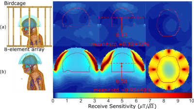 |
94 | Water-Immersed 8-Dipole Receive Array for MR-Guided Focused Ultrasound of the Brain at 3T
Adam Mitchell Maunder1,2, Samuel Pichardo3, G. Bruce Pike3, Melany Mclean3, Fraser Robb4, Ashwin Iyer2, and Nicola De Zanche1,5
1Oncology, University of Alberta, Edmonton, AB, Canada, 2Electrical and Computer Engineering, University of Alberta, Edmonton, AB, Canada, 3Departments of Radiology and Clinical Neurosciences, Hotchkiss Brain Institute, University of Calgary, Calgary, AB, Canada, 4GE Healthcare Inc., Aurora, OH, United States, 5Medical Physics, Cross Cancer Institute, Edmonton, AB, Canada
The piezoelectric transducer array in magnetic resonance guided focused ultrasound shields the RF body-sized birdcage (BC) fields, leading to regions of low receive sensitivity that hinder MR thermometry and structural imaging. This work presents a flexible, acoustically transparent receive array consisting of dipole sections that mimic loops and are contained within the water bath of the ultrasound transducer array helmet surrounding the head. Adjacent elements are decoupled by shared current paths and lumped inductors. In simulation, the receive sensitivity is increased by 7.8-fold compared to a BC and causes a negligible alteration of the transmit field and specific absorption rate.
|
||
1088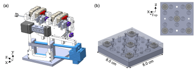 |
95 | Robust Fiducial-based Registration for Mechatronics-Assisted MRI-guided Focal Laser Ablation of Localized Prostate Cancer
Eric Knull1,2, Claire Park2,3, Jeffrey Bax2, David Tessier2, and Aaron Fenster1,2,3
1Biomedical Engineering, Western University, London, ON, Canada, 2Robarts Research Institute, London, ON, Canada, 3Medical Biophysics, Western University, London, ON, Canada
MRI-guided focal laser ablation (FLA) is a promising minimally-invasive therapy method for men with localized prostate cancer. We previously developed an MR-compatible mechatronic system capable of transperineal needle delivery within an in-bore 3T MRI environment. This work presents an improved multi-fiducial structure for robust registration of the mechatronic system for MRI-guided FLA needle delivery. Real-time MRI-guided needle delivery was performed in a tissue-mimicking prostate phantom to virtual targets simulating focal ablation zones. With the implementation of the improved multi-fiducial structure, mechatronics-assisted MRI-guided needle delivery enables a 1.44 mm ablation radius, showing potential utility for accurate FLA to small prostate lesions.
|
||
1089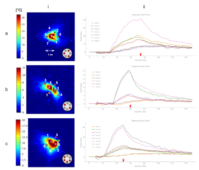 |
96 | A multi-directional laser ablation device for 3D conformational ablation guided by real-time volumetric MR-thermometry
Manon Desclides1,2,3, Guillaume Machinet4, Christophe Pierre4, Valéry Ozenne1,3,5,6, Stéphane Chemouny2, and Bruno Quesson3,5,6
1UMR5536 CRMSB, Université de Bordeaux, Bordeaux, France, 2Certis Therapeutics, Pessac, France, 3IHU Liryc, Electrophysiology and Heart Modeling Institute, Hopital Xavier Arnozan, Pessac, France, 4ALPhANOV, Talence, France, 5Centre de Recherche Cardio-Thoracique de Bordeaux, U1045, University of Bordeaux, Bordeaux, France, 6INSERM, Centre de Recherche Cardio-Thoracique de Bordeaux, U1045, Bordeaux, France
We present here a new laser device allowing us to induce local temperature increase in several directions around the inserted probe to create conformational thermal ablation. The device incorporates 6 independent emitters illuminating several angles radially to the probe. We illustrate the capabilities of the device to create various heating shapes that can be visualized by real-time 2D multi-slice MR-thermometry.
|
||
1090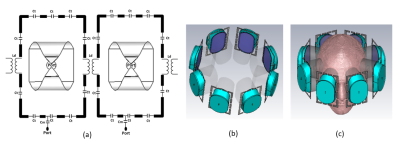 |
97 | 16-Channel Hybrid Dipole+Loop RF Applicator for Thermal MR at 7.0 Tesla
Nandita Saha1, Andre Kuehne2,3, Thomas Wilhelm Eigentler1,4, and Thoralf Niendorf1,2,5
1Berlin Ultrahigh Field Facility (B.U.F.F.), Max-Delbrück Center for Molecular Medicine in the Helmholtz Association, Berlin, Germany, 2MRI.TOOLS GmbH, Berlin, Germany, 3MT MedTech Engineering GmbH, Berlin, Germany, 4Technische Universität Berlin, Chair of Medical Engineering, Berlin, Germany, 5Experimental and Clinical Research Center (ECRC), a joint cooperation between the Charité Medical Faculty and the Max-Delbrück Center for Molecular Medicine in the Helmholtz Association, Berlin, Germany
This work proposes the concept of a loop and SGBT dipole antenna array for diagnostic proton (1H) imaging and thermal intervention at 7.0T MRI. Dipole antennas are the general choice for RF induced heating application as they can be made to operate over a broadband frequency range, have symmetric B1+ field and provide a central E-field distribution. Adding the inherently orthogonal and thus well-decoupled electromagnetic (EM) fields of loops to the dipoles provides additional degrees of freedom for RF shimming and thermal intervention not afforded by only using dipole antennas.
|
||
1091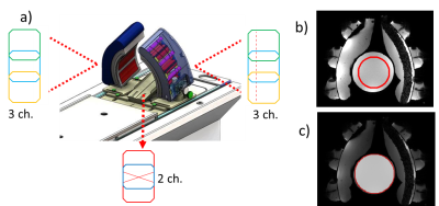 |
98 | Towards motion robust MRT during Microwave Hyperthermia by integrating an 8-channel receiver coil array into the MRcollar
Kemal Sumser1, Gennaro G Bellizzi1, Juan A Hernandez-Tamames2, Gerard C van Rhoon1, and Margarethus M Paulides1,3
1Department of Radiotherapy, Erasmus Medical Center Rotterdam, Rotterdam, Netherlands, 2Department of Radiology and Nuclear Medicine, Erasmus Medical Center Rotterdam, Rotterdam, Netherlands, 3Department of Electrical Engineering, Eindhoven University of Technology, Eindhoven, Netherlands
In the head and neck region, precise heating and temperature monitoring is a challenge. The MRcollar, an MR compatible head and neck microwave hyperthermia applicator has been developed for conformal heating and to enable MR thermometry (MRT) during the treatment. To deliver on the needs of accurate temperature monitoring, the MRcollar has been equipped with 8-channel receiver coil array. This coil array improves increased the SNR by 5 times compared to the body coil of the MRcollar. Increase in the SNR also led to improvement in the MRT precision, from 0.91 °C to 0.37 °C.
|
||
1092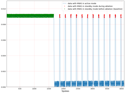 |
99 | 3D stack-of-stars MR thermometry sequence during hepatic microwave ablation: a phantom study with a commercially available microwave generator
Dominik Horstmann1, Bennet Hensen1, Karen Meyer zu Hartlage1, Daniel Luca Reimert1, Josef Joaquin Löning Caballero1, Frank Wacker1, and Marcel Gutberlet1
1Hannover Medical School, Hannover, Germany
A 3D stack-of-stars MR-thermometry sequence was developed and evaluated during hepatic microwave ablation in a chicken phantom. A commercially available microwave generator (MWG) working in pulsed mode controlled by a temperature sensor was used. Results show a spatially dependent phase-offset in the MR-image only during active ablation mode of the pulsed MWG impairing thermometry. Consequently, 73% of thermometry data were discarded for the fixed phantom yielding a temperature precision of 0.72°C±0.60°C whereas 94% were discarded for the moving phantom with an accuracy of 1.08°C±1.25°C. For clinical MR-thermometry, an MWG with less EMI is required to improve resolution and temperature precision.
|
||
1093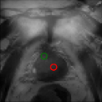 |
100 | Application of Haralick Texture Analysis to Differentiate Suspicious Prostate Lesions from Normative Tissue on Low-field MRI
Dang Bich Thuy Le1, Ram Narayanan1, Meredith Sadinski1, Kathryn Nicholas2, Aleksandar Nacev1, Dinesh Kumar1, and Srirama Venkataraman1
1Promaxo, Oakland, CA, United States, 2Lakeland Radiologists PA, Jackson, MS, United States
Haralick texture features extract frequencies of local spatial variations in signal intensity, quantifying pixel relationships within regions of interest. In this study, Haralick texture features were used to differentiate biopsy-proven, cancerous lesions from non-suspicious regions in low-field MR images. The results demonstrate consistency in texture measures for cancerous regions compared to non-suspicious, where Energy and Homogeneity are elevated while Contrast and Correlation are reduced within cancerous regions compared to non-suspicious regions.
|
||
1094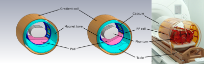 |
101 | Construction of a Tx/Rx body coil on a rotatable patient capsule for MR-guided particle therapy
Kilian A. Dietrich1,2,3, Sebastian Klüter2,4,5, Jürgen Debus1,2,3,4,5,6,7,8, Fabian Dinkel9, Gernot Echner9, Mark E. Ladd1,3,8, and Tanja Platt1,2
1Medical Physics in Radiology, German Cancer Research Center (DKFZ), Heidelberg, Germany, 2Department of Radiation Oncology, Heidelberg University, Heidelberg, Germany, 3Faculty of Physics, Heidelberg University, Heidelberg, Germany, 4National Center for Radiation Research in Oncology (NCRO), Heidelberg, Germany, 5Heidelberg Institute of Radiation Oncology (HIRO), Heidelberg, Germany, 6National Center for Tumor Diseases (NCT), Heidelberg, Germany, 7German Cancer Consortium (DKTK), Heidelberg, Germany, 8Faculty of Medicine, Heidelberg University, Heidelberg, Germany, 9German Cancer Research Center (DKFZ), Heidelberg, Germany
An RF body coil compatible with particle therapy was built for a clinical MR scanner at 1.5T with a rotatable patient capsule. The attenuation of 1H+ and 12C6+ ions due to inelastic scattering was calculated for different materials to estimate the detrimental effects of the RF coil on the particle beam. The imaging capabilities could be demonstrated with phantom measurements at different flip angles, and corresponding transmit and receive characteristics were analyzed and compared to electromagnetic field simulations for both a horizontal and a tilted position of a phantom.
|
||
1095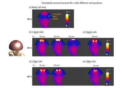 |
102 | Acoustically-transparent passive RF field repeater elements for the mitigation of the low-signal artifact and improved sensitivity in MRgFUS
Isabellle Saniour1, Victor Taracila2, Fraser J.L. Robb2, Rena Fukuda1, Jana Vincent2, Henning U. Voss1, Michael G. Kaplitt3, J. Levi Chazen1, and Simone Angela Winkler 1
1Department of Radiology, Weill Cornell Medicine, New York, NY, United States, 2GE Healthcare, Aurora, OH, United States, 3Department of Neurological Surgery, Weill Cornell Medicine, New York, NY, United States
MRgFUS image quality remains poor and suffers from a low-signal band artifact, preventing efficient image acquisition. We propose to place overlapped acoustically transparent loop elements in “passive mode” between the transducer and the head that act as a transmit field repeater/concentrator. Simulation results not only show mitigation of the low-signal band artifact in the thalamus region, but also demonstrate improvement of the RF transmit signal by a factor of 5 and 2 in the upper brain and the thalamus region, respectively.
|
||
 Back to Meeting Home
 Back to the Program-at-a-Glance
The International Society for Magnetic Resonance in Medicine is accredited by the Accreditation Council for Continuing Medical Education to provide continuing medical education for physicians.
 Back to Meeting Home
 Back to the Program-at-a-Glance
 View the Presentation