Digital Poster
Advanced Liver Imaging Techniques
Joint Annual Meeting ISMRM-ESMRMB & ISMRT 31st Annual Meeting • 07-12 May 2022 • London, UK

Digital Poster
Advanced Liver Imaging Techniques
| Computer # | ||||
|---|---|---|---|---|
2284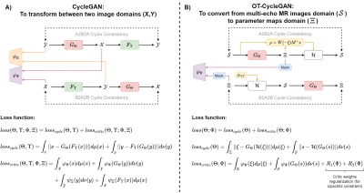 |
20 | Optimal Transport driven Cycle-consistent Generative Adversarial Network (OT-CycleGAN) for an accurate MR water-fat separation
Juan Pablo Meneses1,2,3, Cristobal Arrieta1,2, Gabriel della Maggiora1,2, Pablo Irarrazaval1,2,3,4, Cristian Tejos1,2,3, Marcelo Andia1,2,5, Sergio Uribe1,2,5, and Carlos Sing Long1,2,4,6,7
1Biomedical Imaging Center, Pontificia Universidad Católica de Chile, Santiago, Chile, 2ANID – Millennium Science Initiative Program – Millennium Nucleus for Cardiovascular Magnetic Resonance, Santiago, Chile, 3Electrical Engineering, Pontificia Universidad Católica de Chile, Santiago, Chile, 4Institute for Biological and Medical Engineering, Pontificia Universidad Catolica de Chile, Santiago, Chile, 5Radiology Department, School of Medicine, Pontificia Universidad Catolica de Chile, Santiago, Chile, 6Institute for Mathematical & Computational Engineering, Pontificia Universidad Catolica de Chile, Santiago, Chile, 7ANID – Millennium Science Initiative Program – Millennium Nucleus for Discovery of Structure in Complex Data, Santiago, Chile
MRI water-fat separation is an ill-posed inverse problem usually addressed using complex numerical methods. This problem is related to an MR signal physical model that includes the effects of R2* signal decay ratio and main magnetic field inhomogeneities (Δf), which induce non-linearities that are related to the ill-posedness of the inverse problem. In this work, we propose an Optimal Transport driven Cycle-consistent Generative Adversarial Networks (OT-CycleGAN) framework, which is physics-based, and could use partially labeled training data to estimate the non-linear components (R2* and Δf), and posteriorly compute the water-fat images through a conventional least-squares approach.
|
||
2285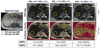 |
21 | Confidence Maps for Reliable Proton Density Fat-Fraction Estimation in the Presence of Low SNR
Jayse M Weaver1,2, Nathan T Roberts2,3, Diego Hernando1,2, and Scott B Reeder1,2,4,5,6
1Medical Physics, University of Wisconsin-Madison, Madison, WI, United States, 2Radiology, University of Wisconsin-Madison, Madison, WI, United States, 3Electrical Engineering, University of Wisconsin-Madison, Madison, WI, United States, 4Biomedical Engineering, University of Wisconsin-Madison, Madison, WI, United States, 5Medicine, University of Wisconsin-Madison, Madison, WI, United States, 6Emergency Medicine, University of Wisconsin-Madison, Madison, WI, United States
Chemical-shift-encoded MRI (CSE-MRI) is an established technique to estimate proton density fat-fraction (PDFF) as a quantitative imaging biomarker of hepatic steatosis. However, due in part to low flip angle acquisitions, CSE-MRI acquisitions often have low SNR leading to biased and noisy PDFF measurements. Unreliable PDFF measurements are particularly problematic considering clinically relevant PDFF thresholds may be as low as 3-5%. There are currently no methods to gauge the reliability of PDFF measurements. We propose a method for generating color-coded CSE-MRI estimation confidence maps, using a normalized residual metric to determine PDFF measurement reliability.
|
||
2286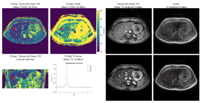 |
22 | Isotropic resolution volumetric liver T2 mapping using a free-breathing navigator-gated radial stack-of-stars T2-prepared Dixon acquisition
Mark Zamskiy1, Kilian Weiss2, Felix N. Harder1, Stefan Ruschke1, Marcus R. Makowski1, Rickmer F. Braren1, and Dimitrios C. Karampinos1
1Department of Diagnostic and Interventional Radiology, School of Medicine, Technical University of Munich, Munich, Germany, 2Philips GmbH Market DACH, Hamburg, Germany
Volumetric liver T2-mapping is of interest in the characterization of focal lesions and diffuse disease but remains technically challenging due to respiratory motion. The present work introduces a novel T2-prepared (T2prep) radial stack-of-stars two-point Dixon gradient echo sequence (T2prep-SoS-Dixon-TFE) for achieving motion-robust 3D isotropic T2-mapping in navigator-gated scans using a B0/B1-insusceptible modified adiabatic BIR-4 RF-pulse for T2prep. T2-mapping is performed via dictionary matching to Bloch simulations for the Dixon-decomposed water signal. The proposed method enables motion-robust liver fat confounder-free volumetric T2 quantification in better agreement with MRS compared to GraSE and may improve the clinical applicability of free-breathing abdominal T2-mapping.
|
||
2287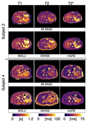 |
23 | Simultaneous T1, T2, and T2* Mapping of the Liver in a Single Breath Hold Using Multi Inversion Spin and Gradient Echo
Mary Kate Manhard1,2, Amol S. Pednekar1,2, Hui Wang1,2,3, Jean A. Tkach1,2, and Jonathan R. Dillman1,2
1Department of Radiology, Cincinnati Children's Hospital Medical Center, Cincinnati, OH, United States, 2Department of Radiology, University of Cincinnati College of Medicine, Cincinnati, OH, United States, 3Clinical Science, Philips Healthcare, Cincinnati, OH, United States Parametric mapping aids in the non-invasive evaluation and longitudinal monitoring of diffuse chronic liver disease. Commercially available parametric mapping techniques require multiple scans over multiple breath-holds. A multi-inversion spin and gradient echo (MI-SAGE) method is presented to simultaneously estimate T1, T2, and T2* at six slice locations in the liver in a single 16s breath-hold. In five volunteers, the MI-SAGE method produced parametric values comparable to those obtained using modified Lock Locker (T1), multiple gradient and spin echo (T2), and multiple gradient echo (T2*) sequences. MI-SAGE offers the potential to significantly shorten the duration of the quantitative MR liver exams. |
||
2288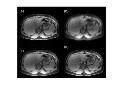 |
24 | Variable density Poisson disc acquisition with iterative deep learning reconstruction for highly accelerated 3D T1-weighted abdominal imaging
Ty A Cashen1, Sangtae Ahn2, Uri Wollner3, Graeme McKinnon4, Isabelle Heukensfeldt Jansen2, Rafi Brada3, Dan Rettmann5, Xucheng Zhu6, and Ersin Bayram7
1GE Healthcare, Madison, WI, United States, 2GE Research, Niskayuna, NY, United States, 3GE Research, Herzliya, Israel, 4GE Healthcare, Waukesha, WI, United States, 5GE Healthcare, Rochester, MN, United States, 6GE Healthcare, Menlo Park, CA, United States, 7GE Healthcare, Houston, TX, United States
3D T1-weighted gradient echo imaging is a key component of the MRI assessment of the abdomen, particularly for the identification and characterization of liver tumors, however, significant acceleration is necessary to consistently mitigate respiratory motion artifact. A variable density Poisson disc undersampled acquisition with a densely connected iterative deep convolutional neural network reconstruction was developed to provide next-generation acceleration up to a factor of 10. On retrospectively undersampled data, the technique outperformed compressed sensing reconstruction in terms of normalized mean-squared error and structural similarity; with a prospectively undersampled scan, the technique maintained image quality in terms of artifact and contrast.
|
||
2289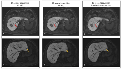 |
25 | Advanced reconstruction provides improved image quality and enables shorter breath-holds in contrast-enhanced liver imaging
David Stockton1, Mihaela Rata1,2, Francesca Castagnoli1, Joshua Shur1, Georgina Hopkinson1, Alison Macdonald1, Marcel Dominik Nickel3, Stephan Kannengiesser3, Dow-Mu Koh1,2, and Jessica M Winfield1,2
1MRI Unit, The Royal Marsden NHS Foundation Trust, London, United Kingdom, 2Division of Radiotherapy and Imaging, The Institute of Cancer Research, London, United Kingdom, 3MR Application Predevelopment, Siemens Healthcare GmbH, Erlangen, Germany
An advanced reconstruction method, which employs a neural network trained on high resolution data to conduct interpolation, combined with iterative denoising, was applied in contrast-enhanced liver imaging in patients receiving a hepatocyte-specific contrast agent. The advanced reconstruction provides improved image quality, with better contrast-to-noise ratio, vessel edge sharpness, and lesion edge sharpness compared with the standard reconstruction using zero-filling interpolation without advanced reconstruction. The improvement in image quality was also observed in faster acquisitions (lower phase resolution), thus enabling shorter breath-hold acquisitions.
|
||
2290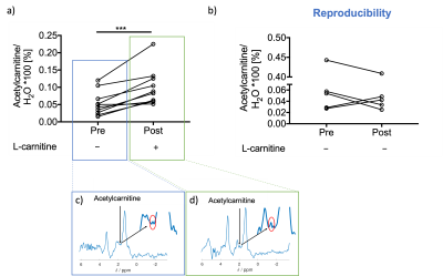 |
26 | L-carnitine Supplementation used as a Tool for Detecting Hepatic Flexibility of Acetylcarnitine using 1H Magnetic Resonance Spectroscopy.
Dragana Savic1, Ferenc E. Mózes1, Peregrine G. Green1, Matthew K. Burrage1, Gabrielle Allen2, Timothy James2, Leanne Hodson1, Stefan Neubauer1, Michael Pavlides1, and Ladislav Valkovič1
1University of Oxford, Oxford, United Kingdom, 2Oxford University Hospitals, Oxford, United Kingdom
This study investigated the in vivo effects on acetylcarnitine levels in the liver after an injection of L-carnitine. L-carnitine facilitates transport of fatty acids into the mitochondria. We show that a single injection of L-carnitine modulated acetylcarnitine levels in the liver and that it modulated several blood markers in the healthy volunteer. Further studies are needed to understand the plasma metabolic changes observed with L-carnitine supplementation.
|
||
2291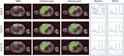 |
27 | Test-retest reliability of in-vivo 31P MRSI of the whole human liver at 7 Tesla using a 31P whole-body transmit coil and 16-channel receive array
Lieke van den Wildenberg1, Ayhan Gursan1, Leonard W.F. Seelen1, Tijl A. van der Velden1, Mark Gosselink1, Martijn Froeling1, Wybe J.M. van der Kemp1, Dennis Klomp1, and Jeanine Prompers1
1Imaging and Oncology, UMC Utrecht, Utrecht, Netherlands
Alterations in 31P metabolite concentrations in the liver can be a consequence of liver disease and therefore 3D 31P MRSI can help in diagnosis and following progression and treatment response. However, commonly used 31P surface coils have a limited penetration depth and do not provide full coverage of the liver. We used an integrated 31P whole-body transmit coil with a local 31P receive array for 31P MRSI of the liver at a 7T MRI scanner. Here we demonstrate that 31P signals can be measured throughout the liver and 31P metabolite levels can be quantified with good to excellent test-retest reliability.
|
||
2292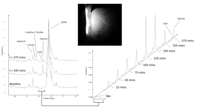 |
28 | Development of 13C MRS measurements of hepatic glutathione production: monitoring oxidative stress in vivo
Stephen Bawden1,2, Bernard Lanz3, Peter E Thelwall4, Guruprasad P Aithal2, and Penny Gowland1
1Sir Peter Mansfield Imaging Centre, School of Physics and Astronomy, University of Nottingham, Nottingham, United Kingdom, 2NIHR Nottingham Biomedical Research Centre, Nottingham University hospitals NHS trust and the University of Nottingham, University of Nottingham, Nottingham, United Kingdom, 3Sir Peter Mansfield Imaging Centre, School of Medicine, University of Nottingham, Nottingham, United Kingdom, 4Translational and Clinical Research Institute, Newcastle University, Newcastle, United Kingdom
Oxidative stress plays a central role in the development of both acute and chronic liver injury, with glutathione being the primary anti-oxidant. This study develops a method of measuring glycine to glutathione flux in vivo using 13C MRS and a novel labelling strategy. Quantification of [2-13C] glycine and [2-13C] glutathione concentrations following glycine ingestion show that the protocol used in this study does successfully enrich the glutathione pool and confirm the assumptions of the metabolic model proposed. This provides a powerful methodology to investigate oxidative stress in patient populations and in response to drug intervention.
|
||
2293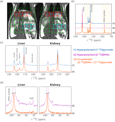 |
29 | Simultaneous Assessment of Complementary Metabolic Pathways in Liver Using Co-polarized Hyperpolarized 13C pyruvate and 13C dihydroxyacetone
Yaewon Kim1, Cornelius von Morze2, Jeremy W. Gordon1, Peder E. Z. Larson1, and Michael A. Ohliger1
1Department of Radiology and Biomedical Imaging, University of California, San Francisco, CA, United States, 2Mallinckrodt Institute of Radiology, Washington University, St. Louis, MO, United States Hyperpolarized 13C MRS is a powerful emerging technique for assessing metabolic liver diseases, including diabetes and fatty liver. Chemical shift separation allows monitoring of many metabolites at once. Here, we explore the possibility of co-polarizing [1-13C]pyruvate and [2-13C]dihydroxyacetone to simultaneously assess two important metabolic pathways that are altered in liver disease. Co-polarization was successful, with preserved T1 relaxation times. Although co-polarization led to modest decreases in polarization levels, all expected metabolites were observed when injected into a healthy rat. By examining two pathways at the same time, co-polarization can be an important tool for assessing liver disease.
|
||
2294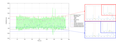 |
30 | Investigating the effects of free breathing on in vivo 13C MRS in the liver at 3T
Abi Spicer1, Susan Francis1, Penny Anne Gowland1, and Stephen Bawden1,2
1Physics, University of Nottingham, Nottingham, United Kingdom, 2NIHR Nottingham Biomedical Research Centre, Nottingham University hospitals NHS trust, Nottingham, United Kingdom
13C Magnetic Resonance Spectroscopy (MRS) in the liver provides the only non-invasive method of studying metabolites in vivo, with standard protocols using multiple averages during a free breathing period. Using a respiratory belt to monitor the breathing cycle and in-house scripts, spectra were binned into inhale and exhale groups categorised as spectra within 2 standard deviations of the average minima/maxima. A systematic reduction in B0 during exhale compared with inhale but minimal impact on average B1 values during an inhale versus exhale. However, no significant changes in Phase angle, area under the curve or FWHM between the data sets.
|
||
2295 |
31 | Combining Convolutional Neural Networks, Multiparametric MRI, and Error Detection to Improve Automated Liver Segmentation Video Permission Withheld
Matthew Gibbons1, Edgar Castellanos Diaz1, Suneil K Koliwad2, Peter W Hunt2, Jean-Marc Schwarz2,3, Kathleen Mulligan2,3, Robert H Lustig4, Alejandro Gugliucci3, Diana L Alba2, Ayca Erkin-Cakmak4, and Susan M Noworolski1
1Department of Radiology and Biomedical Imaging, University of California San Francisco, San Francisco, CA, United States, 2Department of Medicine, University of California San Francisco, San Francisco, CA, United States, 3College of Osteopathic Medicine, Touro University - California, Vallejo, CA, United States, 4Department of Pediatrics, University of California San Francisco, San Francisco, CA, United States
The objective of this study was to generate an automatic liver segmentation method. Two methods were compared. The first, M1, was a Convolutional Neural Network (CNN) trained on proton density fat fraction (PDFF) maps. The second, M2, was the CNN trained on multiparametric MRI (mpMRI) images combined with an error detection protocol. The distributions for Dice similarity coefficient (DSC), volume, and PDFF were improved for M2 versus M1. The DSC mean increased from 0.91 to 0.96. The M2 method was effective in detecting and correcting poor segmentations while significantly reducing processing time as compared to manual segmentation.
|
||
2296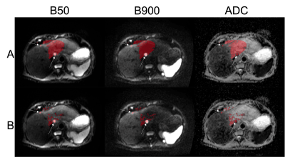 |
32 | Segmentation of Liver Lesions in WB-DWI Using Deformable Registration and Machine Learning Techniques
Annemarie Knill1,2, Antonio Candito1, Jessica Winfield1,2, James Larkin1,2, Samra Turajlic2,3, Dow Mu Koh1,2, Christina Messiou1,2, and Matthew Blackledge1
1The Institute of Cancer Research, London, United Kingdom, 2The Royal Marsden NHS Foundation Trust, London, United Kingdom, 3The Francis Crick Institute, London, United Kingdom We present a proof-of-concept study to assess whether deformable registration followed by tissue classification using machine learning (ML) is an effective method for the delineation of liver metastases in whole-body diffusion-weighted imaging (WB-DWI). Deformable atlas-based registration achieves good quality delineation of the liver (Dice coefficient > 70%) and out of three ML models random forest achieved the best F-1 measure for segmenting disease within the liver. |
||
 Back to Meeting Home
 Back to the Program-at-a-Glance
The International Society for Magnetic Resonance in Medicine is accredited by the Accreditation Council for Continuing Medical Education to provide continuing medical education for physicians.
 Back to Meeting Home
 Back to the Program-at-a-Glance
 View the Presentation