Online Gather.town Pitches
Diagnosis of Neurological Disorders
Joint Annual Meeting ISMRM-ESMRMB & ISMRT 31st Annual Meeting • 07-12 May 2022 • London, UK

| Booth # | ||||
|---|---|---|---|---|
3113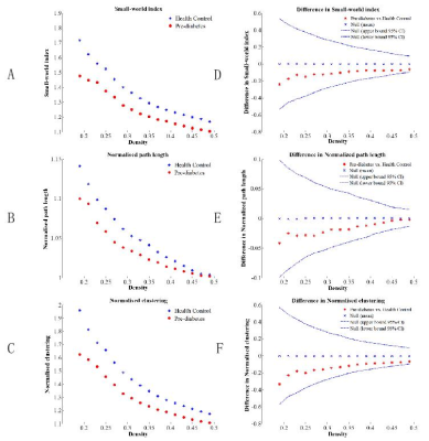 |
1 | Altered segregation of gray matter structural Covariance Networks in prediabetes
Lingling Deng1, Huasheng Liu1, Wen Liu1, Yunjie Liao1, Qi Liang1, Chen Thomas Zhao2, and Wei Wang1
1Department of Radiology, the Third Xiangya Hospital, Central South University, Changsha, China, 2Philips Healthcare, Guangzhou, China
The structural covariance of connected gray matter has been demonstrated valuable in inferring large-scale structural brain networks. The alterations of grey matter structural covariance networks in prediabetes remains unclear. In this study, the topological features and robustness of gray matter structural covariance networks in prediabetes were examined. Results showed that the prediabetes group retained the small-worldness characteristics. The prediabetes group showed higher clustering coefficient, higher local efficiency and more vulnerable to random failure than healthy controls (HCs) group, suggesting that prediabetes disturbed the segregation of gray matter structural covariance networks, which provided new insights into the pathophysiology of this disease
|
||
3114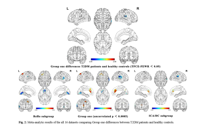 |
2 | Changes of brain function in patients with type 2 diabetes by different analysis methods: A new coordinate based meta-analysis of neuroimaging
Zeyang Li1, Teng Ma1, Lin-Feng Yan1, and Guangbin Cui1
1Hospital, Fourth Military Medical University, 569 Xinsi Road, Xi’an 710038, Shaanxi, China, Xi’an, China
Neuroimaging meta-analysis have identified abnormal neural activity alterations involved in type 2 diabetes mellitus (T2DM) patients, but there is no consistency and heterogeneity analysis between different brain imaging processing strategies. For the indicators obtained from varied post-processing methods reflect different neurophysiological and pathological characteristics, we further conducted a coordinated-based meta-analysis (CBMA) for two categories of neuroimaging literature grouped by similar data processing indicators. Compared to healthy controls, T2DM patients showed a significantly decreased brain activity in the right rolandic operculum, right supramarginal gyrus and right postcentral gyrus, providing a new non-invasive biomarker for T2DM neuropathy.
|
||
3115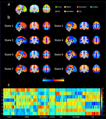 |
3 | Age Related Atypical Dynamic Brain Activity of Autism Spectrum Disorder Analyzed by Co-Activate Pattern
Yunge Zhang1, Dongyue Zhou1, Wei Zhao1, Guoqiang Hu1, Fengyu Cong1, and Huanjie Li1
1School of Biomedical Engineering, Faculty of Electronic and Electrical Engineering, Dalian University of Technology, Dalian, China
Atypical dynamic brain activities (dBAs) of people with autism spectrum disorder (ASD) were reported related to ASD symptoms. However, the most commonly used dynamic function connectivity method is affected by sliding window size. In this study, co-activate pattern analysis which keeps high temporal resolution and without the limitation of window size, is used to evaluate dBAs. It is found that the atypical dBAs of ASD is related to ASD core symptoms, and the atypical dBAs pattern of ASD is also affected by age.
|
||
3116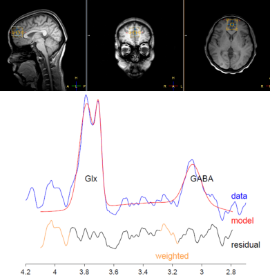 |
4 | MRS study on the correlation between frontal GABA+/Glx ratio and abnormal cognitive function in patients with narcolepsy
Yanan Gao1, Yanting Liu1, Sihui Zhao1, Hui Steve2, Mikkelsen Mark2, Edden A.E. Richard 2, Chen Zhang3, and Bing Yu4
1China Medical University, shenyang, China, 2Russell H. Morgan Department of Radiology and Radiological Science, The Johns Hopkins University School of Medicine, Baltimore, MD, USA F. M. Kirby Center for Functional Brain Imaging, Kennedy Krieger Institute, Baltimore, MD, USA, Baltimore, MD, United States, 3MR Scientific Marketing, Siemens Healthineers, BeiJing, China, 4China Medical University, Shenyang, China
A series of studies have suggested the cognitive deficits in patients with narcolepsy. The aim of the current study is to explore the correlation between abnormal GABA+/Glx ratio in sleep state and abnormal cognitive function in patients with narcolepsy using Hadamard Encoding and Reconstruction of Mega-Edited Spectroscopy (HERMES) method. This study demonstrate that the ratio of GABA+/Glx in prefrontal lobe of narcolepsy patients during N2 sleep stage were higher than that of normal control group, which may be related to their abnormal cognitive functions.
|
||
3117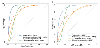 |
5 | Clinical variables, deep learning and radiomics features help predict the prognosis of anti-NMDA receptor encephalitis in Southwest China Video Permission Withheld
Yayun Xiang1, Xiaoxuan Dong2, and Yongmei Li1,3
1Department of Radiology, The First Affiliated Hospital of Chongqing Medical University, Chongqing, China, 2College of Computer & Information Science, Southwest University, Chongqing, China, Chongqing, China, 3The First Affiliated Hospital of Chongqing Medical University, Chongqing, China
The establishment and validation of accurate prognostic models in anti-NMDA receptor (NMDAR) encephalitis is lacking. This study aims to conduct an artifificial intelligence (AI) scheme to predict the prognosis of patients with anti-NMDAR encephalitis using clinical and machine learning features. We first bulid the clinical, deep learning and radiomics models, respectively. Then, we fuse the three schemes to build a fusion model and use an independent external dataset for further validation. The new fusion model significantly outperforms all other models. It demonstrates that applying AI method is an effective way to improve the performance of prognosis prediction in anti-NMDAR encephalitis.
|
||
3118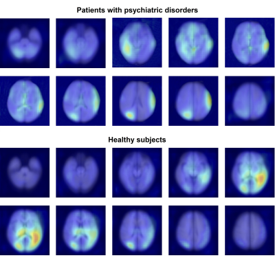 |
6 | Applicable diagnostic model for detecting patients with mental disorders with magnetic resonance imaging
Wenjing Zhang1, Chengmin Yang1, Zehong Cao2, Feng Shi2, and Su Lui1
1Department of Radiology, West China Hospital of Sichuan University, Chengdu, China, 2Department of Research and Development, Shanghai United Imaging Intelligence Co., Ltd., Shanghai, China
To yield clinical utility in mental disorder identification individually, we used a multiple instance learning-based method to construct a digital model based on clinical MRI scans for automated detection of patients with psychiatric disorders. An accuracy of 84% was achieved in the primary dataset with 19453 subjects, and 76% in external dataset with 600 subjects. A higher sensitivity was achieved in identifying high-risk subjects than self-scaled questionnaires (71.1% vs 22.2%) in 148 prospectively recruited college students. With a complete workflow of development and validation, the constructed model is more practical to be translated in high-risk subject screening among vulnerable populations.
|
||
3119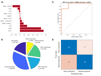 |
7 | Machine learning–based prediction of post-concussive working memory decline: a 1-year fMRI prospective study
Yi-Tien Li1,2, Yung-Chieh Chen2, Yung-Li Chen3, Duen-Pang Kuo2, and Cheng-Yu Chen2
1Neuroscience Research Center, Taipei Medical University, Taipei, Taiwan, 2Translational Imaging Research Center, Taipei Medical University Hospital, Taipei, Taiwan, 3Department of Occupational Therapy, Kaohsiung Medical University, Kaohsiung, Taiwan
The objective of this study is to construct a framework for precise individualized prediction of post-concussive cognitive outcomes based on the early fMRI and neuropsychological biomarkers assessed at baseline to facilitate early therapeutic intervention and individualized rehabilitation strategies. Satisfactory predictions can be achieved for patients whose WM function did not recover after 3 months (accuracy = 87.5%), 6 months (accuracy = 83.3%), and 1 year (accuracy = 83.3%). The results prove the feasibility of using machine learning–based approaches to reveal predictive biomarkers related to poor post-concussive cognitive outcomes.
|
||
3120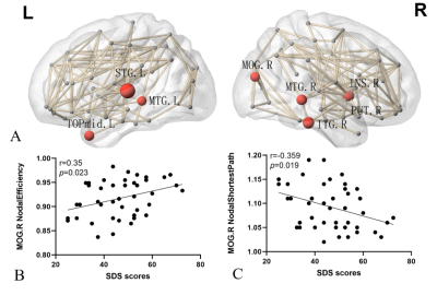 |
8 | Altered brain white matter connectom underlie affective disorders after mild traumatic brain injury at the acute stage
Wenjing Huang1, Jing Zhang1, Wanjun Hu1, Pengfei Zhang1, Jun Wang1, and Shaoyu Wang2
1Lanzhou University Second Hospital, Lanzhou, China, 2MR Scientific Marketing, Siemens Healthineers, ShangHai, China
Mild traumatic brain injury (mTBI) is not a static event, but a progressive injury. The neuroanatomical alterations of mTBI at the acute stage can be an initial step of damage leading to cognitive and emotional deficit. The potential mechanisms conferring these deficits are poorly understood. In this study, we employed graph theory to explore alterations of white matter connectome and to study their relationship with neuropsychological examinations. We found disrupted topologic manner of the brain’s structural connectome, and the changes of right middle occipital gyrus explain depressive symptoms in the acute phase of mTBI due to white matter network alterations.
|
||
3121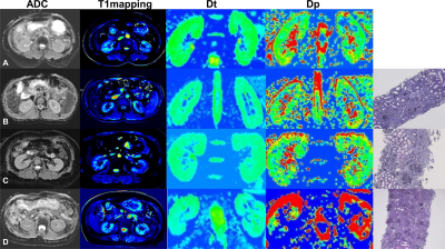 |
9 | The value of multi-parametric magnetic resonance imaging in evaluating renal fibrosis
Chenchen Hua1, Lu Qiu2, Leting Zhou3, Yi Zhuang2, Bin Xu1, Liang Wang3, Min Xu4, and Haoxiang Jiang2
1Diagnostic Radiology, Wuxi People's Hospital Affiliated to Nanjing Medical University, Wuxi, China, 2Diagnostic Radiology, The Affiliated Wuxi Children's Hospital of Nanjing Medical University, Wuxi, China, 3Nephrology, Wuxi People's Hospital Affiliated to Nanjing Medical University, Wuxi, China, 4Human Anatomy, Medical School, Southeast University, Nanjing, China
Chronic kidney disease (CKD) is chronic progressive renal parenchymal damage caused by multiple factors. Evaluation of the degree of renal interstitial fibrosis (IF) is of great importance in treatment and prognosis prediction. Herein, we use multi-parametric magnetic resonance imaging (mpMRI) to assess renal IF noninvasively with intention to identify the optimal MRI biomarkers for clinical application. Our results show that multi-parameter prediction model using cortical longitudinal relaxation time (cT1) and cortical true diffusion coefficient (cDt) can effectively assess the degree of renal IF and support clinical diagnosis, treatment strategy and risk stratification in CKD patients.
|
||
3122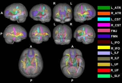 |
10 | Fibre-specific white matter alterations in recovered COVID-19 patients Video Permission Withheld
Yu Shen1, Yan Bai1, Xianchang Zhang2, Yaping Wu1, Menghuan Zhang1, and Meiyun Wang1
1Henan Provincial People's Hospital, Zhengzhou, China, 2MR Collaboration, Siemens Healthineers Ltd, Beijing, China
The brain fibre-specific changes in patients who recovered from Coronavirus Disease 2019 (COVID-19) for one year but still had anosmia (reCOVID19_ano) deserves to be investigated. This study used fixel based analysis (FBA), which is a novel diffusion MRI post-processing technique that enables fibre tract-specific statistical analysis, to investigate the white matter alterations in reCOVID19_ano. The results revealed that the intra-axonal volume in reCOVID19_ano was significantly increased in several fibre tracts. We speculate that impaired fibre tracts may recover gradually in a compensatory mechanism after one year recovery in reCOVID19_ano patients.
|
||
3123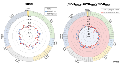 |
11 | Regional comparison of brain standardized uptake values in FBPA PET-MR with FBPA PET-CT
Chien-Ying Lee1,2, Chia-Wei Li3, Chien-Yuan Lin3, Wen-sheng Huang4, and Ko-Han Lin1
1Department of Nuclear Medicine, Taipei Veterans General Hospital, Taipei, Taiwan, 2Department of Biomedical Imaging and Radiological Sciences, National Yang-Ming University, Taipei, Taiwan, 3GE Healthcare, Taipei, Taiwan, 4Department of Nuclear Medicine, Taipei Medical University Hospital, Taipei, Taiwan
18F-fluoro-Boronophenylalanine (FBPA) Positron emission tomography (PET) plays a crucial role for patient selection prior to boron neutron capture therapy. As integrated PET-MR is being used frequently in clinical research and routine, this study aimed to compare regional difference of brain standard uptake values on the PET quantification accuracy in FBPA PET-CT and PET-MR among lesions and normal tissues. Compared to SUVR in PET-CT, PET-MR data showed significant lower SUVR in lesion regions, while no significance was observed in the normal tissue regions. The result of this study may accelerate the routine use of PET-MR in tumoral quantification before BNCT.
|
||
3124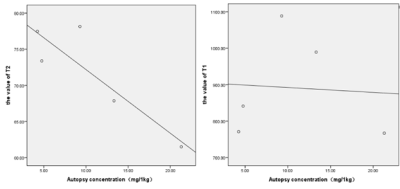 |
12 | The T1mapping, T2mapping findings in patients with minimal hepatic encephalopathy and their impact on cognition value
Wenxiao Liu1,2, Minglei Wang2, Xiaocheng Wei3, Min Li3, Jianguo Zhao2, Xin Ge1, Xuhong Yang1, Peng Yong1, and Xiaodong Wang2
1Ningxia Medical University, Yinchuan, China, 2General Hospital of Ningxia Medical University, Yinchuan, China, 3GE Healthcare, Beijing, China
In this study, we aim to investigate whether T1mapping and T2mapping can be used as diagnostic tools for screening MHE, and strive earlier time for diagnosis and treatment for these patients. It was concluded that T1 values of bilateral putamen, right globus pallidus and bilateral frontal lobe in MHE group were statistically significant compared with HC group and NMHE group and the T2 values of right putamen and right frontal lobe were statistically significant. In addition, the T2 values of the HC group in this study were highly correlated with Hallgren and Sourander’s autopsy results.
|
||
3125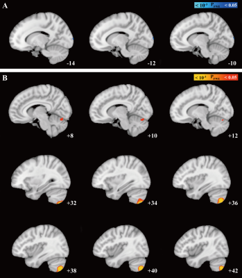 |
13 | Cerebellar iron accumulation in idiopathic cervical dystonia evaluating by quantitative susceptibility mapping Video Not Available
Hongxia Li1, Ming Zhang2, Hongjiang Wei2, and Yiwen Wu1
1Department of Neurology & Institute of Neurology, Ruijin Hospital, affiliated with Shanghai Jiao Tong University School of Medicine, Shanghai, China, 2School of Biomedical Engineering, Shanghai Jiao Tong University, Shanghai, China
We found increased susceptibility in patients compared with HCs that primarily involved cerebellar structures . The result of 2D-flatmap visualization showed that the involved subregions of iron accumulation were in the right Crus Ⅱ and right lobule Ⅶb. Interestingly, no significant differences in QSM value of basal ganglia, thalamus, or motor cortex were observed between ICD patients and HCs. Moreover, there was no significant difference between groups regarding the GMV in the cerebellum. Of consistent findings with standard structural MRI technique highlights the potential of probing tissue iron across the cerebellum with QSM as a disease biomarker
|
||
3126 |
14 | Stage-dependent differential influence of metabolic and structural networks on memory across Alzheimer’s disease continuum Video Permission Withheld
Xing Qian1, Kok Pin Ng2,3,4, Kwun Kei Ng1, Fang Ji1, Pedro Rosa-Neto 5,6, Serge Gauthier 6, Nagaendran Kandiah 2,3,4, and Juan Helen Zhou 1,3,7,8
1Centre for Sleep and Cognition and Centre for Translational MR Research, Yong Loo Lin School of Medicine, National University of Singapore, Singapore, Singapore, Singapore, Singapore, 2Department of Neurology, National Neuroscience Institute, Singapore, Singapore, Singapore, 3Duke-NUS Medical School, Singapore, Singapore, Singapore, 4Lee Kong Chian School of Medicine, Nanyang Technological University Singapore, Singapore, Singapore, Singapore, Singapore, 5Translational Neuroimaging Laboratory, The McGill University Research Centre for Studies in Aging, Montreal, Canada, Montreal, QC, Canada, 6Alzheimer’s Disease Research Unit, The McGill University Research Centre for Studies in Aging, McGill University, Montreal, Canada, Montreal, QC, Canada, 7Department of Electrical and Computer Engineering, National University of Singapore, Singapore, Singapore, Singapore, Singapore, 8Integrative Sciences and Engineering Programme (ISEP), National University of Singapore, Singapore, Singapore, Singapore, Singapore
While emerging evidence suggests the association between network neurodegeneration and memory varies with pathology in Alzheimer’s disease (AD), the trajectory of this relationship remains elusive. We stratified 708 participants into non-amyloid/non-tau, tau-only, and AD pathology groups and examined the associations between individual-level structural and metabolic network integrity and memory across cognitive stages (cognitively normal, mild cognitive impairment, and probable AD) in each pathology group. The associations of hippocampal and default mode networks with memory exhibited differential pathology-dependent trajectories across cognitive stages. Our findings pave the way for early interventions and stage-dependent remedies to modify disease trajectory and improve clinical outcomes.
|
||
3127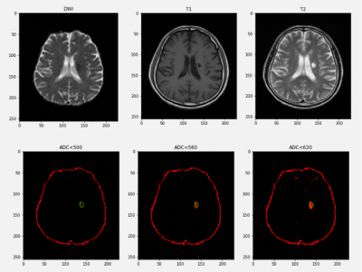 |
15 | Infarct core estimation with ADC: A retrospective study Video Permission Withheld
Yingying Dong1, Yu Luo2, Mingming Wang2, Quan Tao2, and He Wang3
1Institute of Science and Technology for Brain-Inspired Intelligence, Fudan University, Shanghai, China, 2Department of Radiology, Shanghai Fourth People's Hospital, School of Medicine, Tongji University, Shanghai, China, 31Institute of Science and Technology for Brain-Inspired Intelligence, Fudan University 2Human Phenome Institute, Fudan University, Shanghai, China
According to a large amount of emergency and follow-up data, we propose an optimized threshold of the apparent diffusion coefficient to identify the ischemic core with fair accuracy, which is crucial for the decision of the treatment scheme and operation mode.
|
||
The International Society for Magnetic Resonance in Medicine is accredited by the Accreditation Council for Continuing Medical Education to provide continuing medical education for physicians.