Online Gather.town Pitches
Diagnosis of Liver & Kidney Disease
Joint Annual Meeting ISMRM-ESMRMB & ISMRT 31st Annual Meeting • 07-12 May 2022 • London, UK

| Booth # | ||||
|---|---|---|---|---|
4654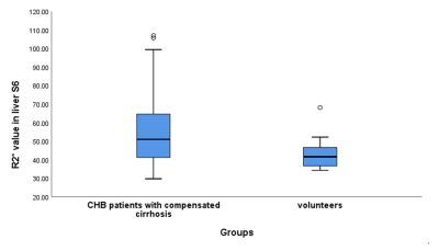 |
1 | Distinction of compensated stage of liver cirrhosis in patients with chronic hepatitis B using IDEAL-IQ Magnetic Resonance Imaging
Hongyi Li1, Huang lesheng1, Jun Chen1, Weiyin Vivian Liu 2, Jinghua Jiang1, Kaili Cai1, Tianzhu Liu1, Wanchun Zhang1, Jiahui Tang1, Kailian Yang1, Se Peng3, and Dan Li4
1Radiology, Guangdong Hospital of Traditional Chinese Medicine, Zhuhai, China, 2MR Research, GE Healthcare, Beijing, China, 3Clinical Lab, Guangdong Hospital of Traditional Chinese Medicine, Zhuhai, China, 4Hepatology, Guangdong Hospital of Traditional Chinese Medicine, Zhuhai, China
The purpose was to evaluate the feasibility of IDEAL-IQ in distinction of patients with early liver cirrhosis(compensated stage). Sixty CHB patients with compensated cirrhosis conformed by liver biopsy and twenty healthy controls were recruited. FF and R2* of IDEAL-IQ sequence were analyzed between CHB compensated cirrhosis patients and volunteers. Correlations between FF and R2* and ALT, INR values were analyzed in CHB patients with compensated cirrhosis. R2* was statistically different between compensated cirrhosis patients and volunteers (p<0.01) with AUC of 0.726. The correlations between FF and ALT, INR were both fair(0.268, p=0.03 and -0.351, p<0.01).
|
||
4655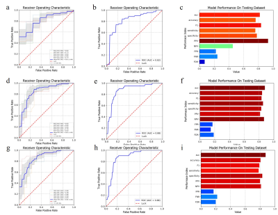 |
2 | Staging liver fibrosis through a machine learning model built from radiomics features of T2WI images
nannan shi1, jianqing sun2, Fei Shan1, Yiying qin1, Zecheng yang1, Weibo chen3, and yuxin shi1
1Shanghai Public Health Clinical Center, Fudan University, shanghai, China, 2Shanghai United Imaging Research Institute of Intelligent Imaging, shanghai, China, 3Philips Healthcare, shanghai, China
To further develop and validate a radiomics-based model from liver T2WI images for staging liver fibrosis.
|
||
4656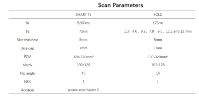 |
3 | Quantitative T1 and R2* mapping in the evaluation of renal function for diabetic nephropathy patients
Huijian Lu1, Hongmei Gu1, Fangfang Shang1, Li Yuan1, Xinquan Wang1, Weiqiang Dou2, and Weiyin Vivian Liu2
1Affiliated Hospital of Nantong University, Nantong, China, 2MR Research, GE Healthcare, Beijing, China
Renal T1 and R2* mapping were performed on 55 diabetic nephropathy (DN) patients , 20 healthy volunteers(HVs)and 10 diabetes mellitus (DM) patients to explore the clinical potential in assessing renal function. While comparable T1 and R2* values of renal cortex were found between HVs and DM patients, DN patients showed significantly increased T1 and R2* values of renal cortex relative to HVs and DM patients. Both T1 and R2*metrics were also significantly correlated with renal function. Therefore, quantitative T1 and R2* mapping might be considered effective measures in evaluating renal function for DN patients.
|
||
4657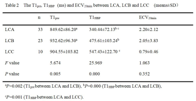 |
4 | Can extracellular volume fraction assess liver fibrosis? A preliminary Gd-EOB-DTPA-enhanced MRI study
Pin Yang1, YanLi Jiang1, Jie Zou1, Miao Chang1, Jing Zhang1, and Kai Ai2
1Department of Magnetic Resonance, LanZhou University Second Hospital, Lanzhou, China, 2Philips Healthcare, Xi’an, China
This study evaluates the feasibility of extracellular volume fraction (ECV) in judging liver fibrosis, analyzes the interaction between ECV and liver function.The method is to calculate ECV in different degrees of liver function and compare the correlation between ECV and serum albumin. Our research shows that ECV20min had no significant difference among Child-Pugh A, Child-Pugh B and Child-Pugh C, there was a weak correlation with serum albumin. Therefore, we consider that ECV of hepatobiliary phase will not be affected by liver function when Gd-EOB-DTPA is used to evaluate liver fibrosis, it may be a reliable tool in assessing liver fibrosis.
|
||
4658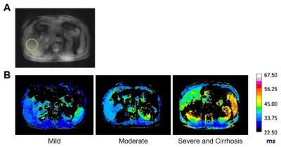 |
5 | Noninvasive Assessment of Chronic Hepatitis B with Different Courses or Liver Cirrhosis Patients Receiving Interferon Therapy Using T1ρ Mapping
Yufei Zhao1 and Xingui Peng1
1Southeast University, Nanjing, China
The staging of chronic hepatitis B mainly relies on invasive pathological biopsy, interferon treatment can reduce the risk of deterioration. 93 patients were divided into three groups: mild, moderate, severe or liver cirrhosis according to histopathology and received T1ρ magnetic resonance liver scan before and after interferon treatment. With the aggravation of chronic hepatitis B, T1ρ relaxation time gradually increased, but it decreased significantly after treatment, which is linearly correlated with the level of ALT and AST. T1ρ magnetic resonance can non-invasively assess the severity of the chronic hepatitis B and monitor the improvement of fibrosis after receiving treatment dynamically.
|
||
4659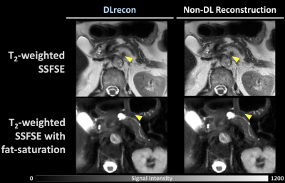 |
6 | Deep Learning Reconstruction for Abdomen Diagnosis: Improvement of Image Quality and Diagnostic Performance
Po-Ting Chen1,2, Cheng-Ya Yeh2, Yi-Chen Chen2, Chia-Wei Li3, Charng-Chyi Shieh3, Chien-Yuan Lin3, and Kao-Lang Liu1,2
1Department of Medical Imaging, National Taiwan University Hospital and National Taiwan University College of Medicine, Taipei, Taiwan, 2Department of Medical Imaging, National Taiwan University Cancer Center, Taipei, Taiwan, 3GE Healthcare, Taipei, Taiwan
There has been no study that demonstrated the quality improvement in abdomen images by DLrecon. We evaluate the image quality comparison, included SNR, CNR, and clinical scoring, between DL and non-DL abdomen images. DLrecon are found to be less artefacts, higher SNR, CNR, and clinical scores than those on non-DL images. This study proves the improvement of image quality by DLrecon, and DLrecon shows its potential power in clinical diagnosis of abdomen images to overcome classical MRI trade-off resolution, SNR, and scan time.
|
||
4660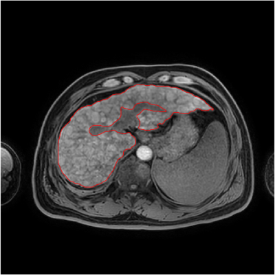 |
7 | Detection of Early Cirrhosis by Means of Curvature Analysis of Liver Surface Nodularity in MRI
Wang Nan1,2,3, Huang Jisui2, Song Qingwei1, Chen Lihua1, Lei Na3, and Liu Ailian1
1the First Affiliated Hospital of Dalian Medical University, Dalian, China, 2Capital Normal University, Beijing, China, 3Dalian University of Technology, Dalian, China
The normal liver is affected by various pathogenic factors, resulting in liver fibrosis, progressive disease, false lobule formation and cirrhosis. Liver cirrhosis will be accompanied by morphological changes of liver. Literature studies have shown that liver surface nodularity can be used to evaluate the progress of liver cirrhosis and predict the stage of liver cirrhosis. The method of curvature analysis of liver surface nodules adopted by our team is feasible and can be used to evaluate early cirrhosis.
|
||
4661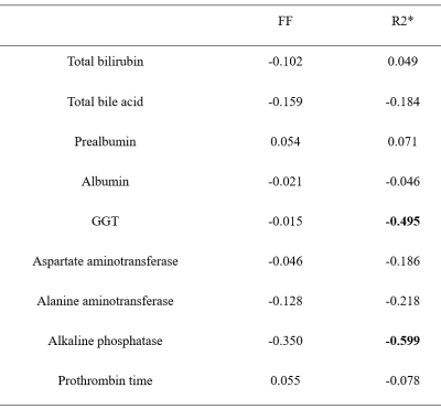 |
8 | Correlation analysis between quantitative parameters of mDIXON-Quant sequence and liver function in patients with liver cirrhosis
Wang Nan1, Lihua Chen1, Qingwei Song1, Jiazheng Wang2, Zhigang Wu2, and Liu Ailian3
1Department of Radiology, the First Affiliated Hospital of Dalian Medical University, Dalian, China, 2Philips Healthcare, Beijing, China, 3the First Affiliated Hospital of Dalian Medical University, Dalian, China
Evaluation of liver reserve function is crucial for patients with cirrhosis and is helpful for the determination of the surgical plan formulation, the evaluation of treatment process and prognosis, and the operation of cirrhosis. However, there is still lacking a clinically useable non-invasive technique to measure the abnormality of liver function. The purpose of this study is to investigate the potential correlation between Fat Fraction (FF), R2* and liver function indexes in patients with cirrhosis using mDIXON-Quant sequence. We found that there was a moderate correlation between R2* value and liver function assay indexes in liver cirrhosis.
|
||
4662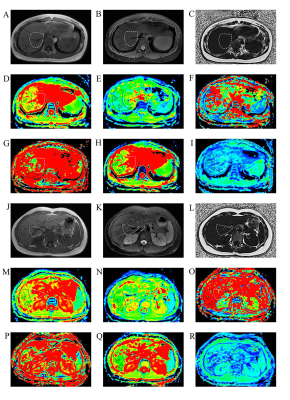 |
9 | Repeatability and reliability of intravoxel incoherent motion derived parameters in evaluation of liver heterogeneity
Tianzhu Liu1, Weiyin Vivian Liu2, Jun Chen1, Lesheng Huang1, Hongyi Li1, Jinghua Jiang1, Kaili Cai1, Jiahui Tang1, Wanchun Zhang1, Kailian Yang1, Guangjun Tian3, Meng Hu3, Se Peng4, Dong Zhang3,
Dong Zhang3, Dan Li3, and Yongxiang Zhuo3
1Radiology, Guangdong Hospital of Traditional Chinese Medicine, Zhuhai, Zhuhai, China, 2MR Research, GE Healthcare, Beijing, China, Beijing, China, 3Hepatology, Guangdong Hospital of Traditional Chinese Medicine, Zhuhai, Zhuhai, China, 4Laboratory, Guangdong Hospital of Traditional Chinese Medicine, Zhuhai, Zhuhai, China
This study aimed to investigate the repeatability and reliability of IVIM derived parameters in evaluation of liver heterogeneity. Patients with early liver fibrosis and healthy subjects were recruited and underwent MRI using respiratory triggered SS-DWI with 12 b values and an IDEAL-IQ sequence. Two readers independently analyzed liver segment data using mono-exponential, bi-exponential and stretched exponential models. CVs and ICCs were calculated before and after the exclusion of subjects with potential liver fat deposition. ADC, D and α measurements of right liver lobe were satisfactorily repeatable, regardless of fat deposition. Segment-VII and -VIII values may be more repeatable and reliable.
|
||
4663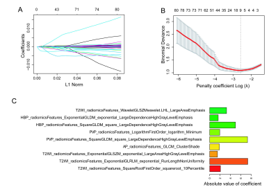 |
10 | Radiomics Analysis of Multi-sequence MR Imaging for Staging Liver Fibrosis and Hepatitis Activity of Chronic Hepatitis B
Aina HUANG1, Jian LU1, Tao ZHANG1, Xueqin ZHANG1, Shuangshuang LU2, and Xiance ZHAO3
1Affiliated Nantong Hospital 3 of Nantong University, Nantong, Jiangsu, China, 2Affiliated Hospital of Nantong University, Nantong,Jiangsu, China, 3Philips Healthcare, Shanghai, China
Synopsis: The purpose of this study was to explore the evaluation value of liver fibrosis staging and hepatitis activity of chronic hepatitis B based on multi-sequence MRI radiomics model. Unenhanced T2WI, T1WI and Gd-EOB-DTPA enhanced T1WI, including AP, PP, TP and HBP, were acquired. And a multi-sequence MRI radiomics model were built. We used receiver operating characteristic (ROC) curves for the diagnostic performance of each sequence and the combination of these six sequences in the training set and validated with the validation set. The models combined with six sequences performed the best.
|
||
4664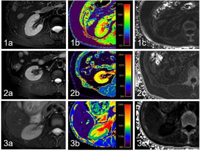 |
11 | Evaluation of T1 and R2* Mapping in Diagnosis of Chronic Kidney Disease
Yue Wang1, Ye Ju1, Liangjie Lin2, and Ailian Liu1
1the First Affiliated Hospital of Dalian Medical University, Dalian, China, 2Philips Healthcare, Beijing, China
Chronic kidney disease (CKD), which has shown increased incidence gradually, is prevalent worldwide and occurs in conjunction with cardiovascular disease and diabetes. Purpose of this study is to evaluate the value of quantitative T1 and R2* mapping in diagnosis of CKD. Results showed that renal cortex R2* and T1 values can be used to distinguish mild CKD from healthy volunteers, and the combination of them can improve the diagnostic efficiency. Cortex R2*, cortex T1, and medulla T1 values can be used to distinguish mild from severe CKD, and the diagnostic efficiency was also significantly improved by using their combination.
|
||
4665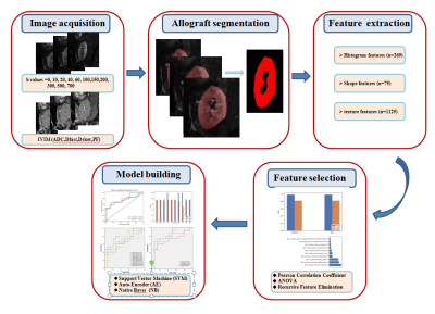 |
12 | Radiomics Analysis Based on IVIM-DWI for Early Assessment of Transplanted Kidney Dysfunction Video Permission Withheld
Xiaodong Liu1, Lihua Chen1, Wen Shen1, Kun Zhang1, Xiaodong Ji1, Robert Grimm2, and Jinxia Zhu3
1Department of Radiology, Tianjin First Central Hospital, Tianjin, China, 2MR Application Predevelopment, Siemens Healthcare GmbH, Erlangen, Germany, 3MR Collaboration, Siemens Healthcare Ltd., Beijing, China
This study investigated the feasibility of predicting early renal function impairment after renal transplantation based on an intravoxel incoherent motion imaging (IVIM)diffusion weighted imaging (DWI)radiomics model. For noninvasive detection of kidney function impairment at an early stage, IVIM-DWI applies a bi-exponential model to evaluate both capillary perfusion and tissue diffusion. The overall accuracy of the radiomics model was 79.3%, with 73.3% sensitivity and 85.7% specificity in distinguishing renal function impairment after renal transplantation. These results demonstrate the potential of a radiomics model for a reliable non-invasive diagnosis of renal transplant function.
|
||
4666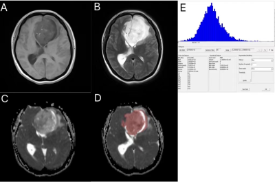 |
13 | Predict MGMT promoter methylation and P53 gene status of glioma based on ADC Histogram Analysis
Huan Zhao1, Yan Bai1, Xianchang Zhang2, and Meiyun Wang1
1Henan Provincial People's Hospital, Zhengzhou, China, 2MR Collaboration,Siemens Healthineers Ltd., Beijing, China
Conventional MRI is limited in reflecting tumor genetic status. This study used the parameters derived from histogram analysis of apparent diffusion coefficient(ADC) maps in glioma lesion to predict the MGMT and P53 gene status. We found the ADC histogram derived parameters are significantly higher in MGHT-methylated and P53 wild group. ROC analysis revealed these parameters have high sensitivity to differentiate the MGHT-methylated/-unmethylated or P53 wild/ mutant gliomas. These findings suggest ADC histogram could be a good candidate for predicting MGMT promoter methylation and P53 gene status in gliomas.
|
||
4667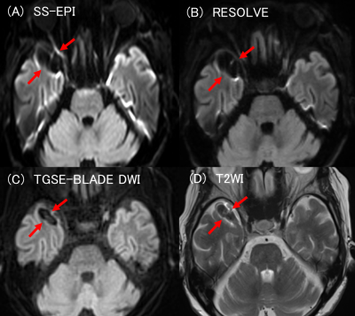 |
14 | Comparison of TGSE-BLADE, RESOLVE and SS-EPI for diffusion weighted imaging after cerebral aneurysmal clipping Video Permission Withheld
Sachi Okuchi1, Yasutaka Fushimi1, Satoshi Nakajima1, Akihiko Sakata1, Takuya Hinoda1, Sayo Otani1, Azusa Sakurama1, Krishna Pandu Wicaksono1, Hiroshi Tagawa1, Yang Wang1, Satoshi Ikeda1, Kun Zhou2, and Yuji Nakamoto1
1Department of Diagnostic Imaging and Nuclear Medicine, Graduate School of Medicine, Kyoto University, Kyoto, Japan, 2Siemens Shenzhen Magnetic Resonance Ltd., Shenzhen, China
TGSE-BLADE DWI has been reported to reduce geometric distortion and susceptibility artifacts. However, it is unknown whether it reduces the clip-induced artifacts and improves image quality in patients after cerebral aneurysmal clipping. We compared the distortion and the artifacts between SS-EPI, RESOLVE, and TGSE-BLADE DWI in healthy volunteers and patients with a cerebral aneurysmal clip. TGSE-BLADE DWI has the best image quality regarding distortion and artifacts, especially at air-bone interfaces and near metal clips, which suggests that TGSE-BLADE DWI is a promising method for evaluating lesions at air-bone interfaces and scanning the patients with a clip.
|
||
The International Society for Magnetic Resonance in Medicine is accredited by the Accreditation Council for Continuing Medical Education to provide continuing medical education for physicians.