Online Gather.town Pitches
Cardiovascular Anatomy, Function, Hemodynamics III
Joint Annual Meeting ISMRM-ESMRMB & ISMRT 31st Annual Meeting • 07-12 May 2022 • London, UK

| Booth # | ||||
|---|---|---|---|---|
4913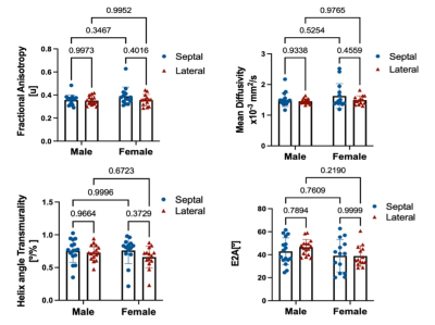 |
1 | Evaluation of Sex and Age Differences in Myocardial Microstructure using Cardiac Diffusion Tensor Imaging
Shi Chen1, Jaume Coll-Font1,2,3, Robert Eder1, Anna Foster 1, Salva Yurista 1,2,3, and Christopher T. Nguyen1,2,3,4
1Cardiovascular Research Center, Massachusetts General Hospital, Charlestown, MA, United States, 2Athinoula A. Martinos Center for Biomedical Imaging, Massachusetts General Hospital, Charlestown, MA, United States, 3Harvard Medical School, Boston, MA, United States, 4Division of Health Science Technology, Harvard-Massachusetts Institute of Technology, Cambridge, MA, United States Sexes and ages are known factors that contribute to cardiovascular pathophysiological variances and prognosis. This study used in-vivo cardiac diffusion tensor MRI (DT-MRI) to characterize how myocardial microstructure differs across sexes and ages. To that purpose, thirty healthy volunteers (14 female) were imaged with in-vivo cardiac DT-MRI and common DT-MRI parameters (MD, FA, HAT, E2A) were computed within the left ventricle (LV). No significant sex-related differences (11%, 9%, 3%, 9% respectively for MD, FA, HAT, E2A at septal regions) or age-related correlations were found. Taken together, sex and age had no effects on the quantification of in-vivo cardiac DT-MRI parameters. |
||
4914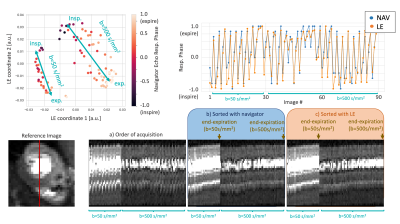 |
2 | Manifold-based Respiratory Phase Estimation Enables Motion and Distortion Correction of Free-Breathing Cardiac Diffusion Tensor MRI
Jaume Coll-Font1,2,3, Shi Chen1, Robert Eder1, Yilin Fan4, Qiao Joyce Han1, Maaike van den Boomen1,2,3, David Sosnovik1,2,3, Choukri Mekkaoui2,3, and Christopher T. Nguyen1,2,3
1Cardiology, Massachusetts General Hospital, Charlestown, MA, United States, 2Harvard Medical School, Boston, MA, United States, 3Athinoula A. Martinos Center for Biomedical Imaging, Massachusetts General Hospital, Charlestown, MA, United States, 4Massachusetts Insitute of Technology, Cambridge, MA, United States
For in-vivo cardiac diffusion-tensor MRI (DT-MRI), respiratory motion and B0 field inhomogeneities produce misalignment and geometric distortion in diffusion weighted images acquired with conventional single shot echo planar imaging. We propose using Laplacian Eigenmaps (LE), a dimensionality reduction method, to retrospectively estimate the respiratory phase of DWI and facilitate both distortion correction (DisCo) and motion compensation (MoCo). LE-based DisCo and MoCo reduces geometric distortion by 13.2% while producing computationally efficient image alignment. Furthermore, preliminary results indicate the LE-based DisCo and MoCo can be applied with only acquiring a single set of b-value averages.
|
||
4915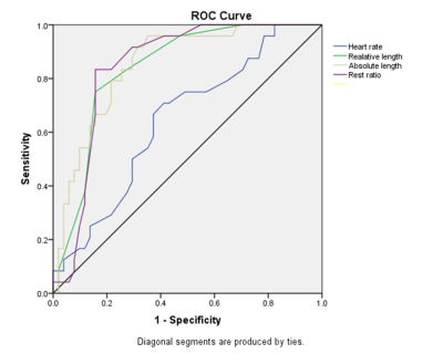 |
3 | Cine MRI detects shorter cardiac rest periods in pulmonary hypertension
Kai Lin1, Roberto Sarnari1, Ashitha Pathrose1, Daniel Gordon1, Michael Markl1, and James Carr1
1Radiology, Northwestern University, Chicago, IL, United States
The existence of pulmonary hypertension (PH) can independently shorten the length of cardiac rest periods. The length of cardiac rest periods can be used to discriminate PH patients from healthy controls.
|
||
4916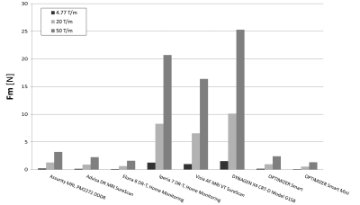 |
4 | A Comparison of the Magnetic Force of MR-Conditional Cardiac Implants
Mabel Shehada1, Emile Shehada2, Ramez Shehada3, and David Prutchi4
1UCSD, San Diego, CA, United States, 2Yale University, New haven, CT, United States, 3Medical Technology Laboratories, La Mirada, CA, United States, 4Impulse Dynamics, MARLTON, NJ, United States
For implant systems composed of an implantable pulse generator (IPG) and lead(s), the MR-Conditional labeling specifies a magnetic spatial gradient for the whole system but not for its individual components. To identify the component experiencing the highest magnetic force, thus dictating the safe limit, we measured multiple IPGs and leads per ASTM F2052-15. The highest force on leads under 4.77T/m, 20T/m, and 50T/m was 0.003N, 0.021N, and 0.053N, respectively. The lowest IPG forces were 0.080N, 0.529N, and 1.322N. For any magnetic spatial gradient, the lowest IPG force was 2,480% greater than the highest lead force.
|
||
4917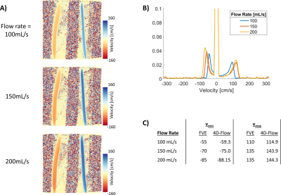 |
5 | Venc Scouting with a Whole Volume Fourier Velocity Encoding Spectrum Acquisition
Michael Loecher1,2 and Daniel B Ennis1,2,3
1Radiology, Stanford University, Stanford, CA, United States, 2Radiology, Veterans Administration Health Care System, Palo Alto, CA, United States, 3Cardiovascular Institute, Stanford University, Stanford, CA, United States We explored the application of acquiring Fourier Velocity Encoding (FVE) without spatial encoding to quickly estimate the velocity distribution within a volume of interest. One application is to allow for quick selection of Venc for a subsequent 4D-Flow acquisition, which can limit velocity aliasing and fine-tune the velocity-to-noise ratio for better data quality. Other applications may include new ways of looking at the pulsatility or turbulence of a volume of interest. In this work we implement a FVE sequence in Pulseq and run it in flow phantoms to demonstrate the proof-of-concept data of this technique. |
||
4918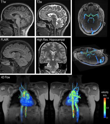 |
6 | Heart-brain MRI evaluation in one exam: a feasibility study in healthy aging adults
Kelly Jarvis1, Jackson E Moore1, Maria Aristova1, Ning Jin2, Valerie Torres1, Susanne Schnell1,3, Eric Russell1, Michael Markl1, Emily Rogalski4, and Ann Ragin1
1Radiology, Northwestern University, Chicago, IL, United States, 2Siemens Medical Solutions Inc., Cleveland, OH, United States, 3Medical Physics, University of Greifswald, Greifswald, Germany, 4Psychiatry and Behavioral Sciences, Northwestern University, Chicago, IL, United States
Mechanisms underlying heart-brain hemodynamic coupling and effects on the brain remain unclear due to challenges of measuring both heart and brain in a single MRI exam. We have developed a comprehensive MRI protocol that incorporates 1) 4D flow MRI of chest, 2) 4D flow MRI of head and 3) structural neuroimaging into one MRI exam to assess cardiovascular and cerebrovascular flow as well as white matter lesions and brain atrophy. This study demonstrates the utility of heart-brain MRI as viable new tool for in vivo evaluation of hemodynamic coupling along the entire heart-brain pathway.
|
||
4919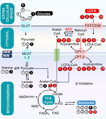 |
7 | Effect of Knockout of Two Acetyl-CoA Carboxylase Isoforms in the Isolated Mouse Heart on Oxidation of Exogenous and Endogenous Energy Sources
Gaurav Sharma1, Chai-Wan Kim2,3, Xiaodong Wen1, A. Dean Sherry1,4,5, Chalermchai Khemtong1,6,7, Craig R. Malloy1,2,4, and Jay D. Horton2,3
1Advanced Imaging Research Center, University of Texas Southwestern Medical Center, Dallas, TX, United States, 2Department of Internal Medicine, University of Texas Southwestern Medical Center, Dallas, TX, United States, 3Department of Molecular Genetics, University of Texas Southwestern Medical Center, Dallas, TX, United States, 4Department of Radiology, University of Texas Southwestern Medical Center, Dallas, TX, United States, 5Department of Chemistry, University of Texas at Dallas, Richardson, TX, United States, 6Department of Medicine, Division of Endocrinology, Diabetes and Metabolism, University of Florida, Gainesville, FL, United States, 7Department of Biochemistry and Molecular Biology, University of Florida, Gainesville, FL, United States
The product of acetyl-CoA carboxylase (ACC), malonyl-CoA, inhibits oxidation of long chain fatty acids by mitochondria. Cardiac-specific deletion of ACC-2 is associated with increased oxidation of fatty acids, as expected, the effects on glucose oxidation are controversial, and increased oxidation of stored triglycerides has been postulated. Expression of the other isoform, ACC-1, is preserved in ACC-2 mutant hearts, so alternative sources of malonyl-CoA may be important. We found that knock out of both isoforms was associated with a small increase in fatty acid oxidation, a small decrease in glucose oxidation, and little effect on oxidation of stored energy supplies.
|
||
4920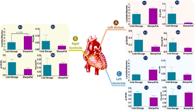 |
8 | Preservation of Human Hearts for Transplantation: Comparison of Metabolic Indicators after Conventional and Temperature Controlled Storage
Gaurav Sharma1, Ryan J. Vela2, LaShondra Powell2, Stanislaw Deja3,4, Monika Mizerska3, Michael E. Jessen2, Shawn C. Burgess3,5, Craig R. Malloy1,6,7, and Matthias Peltz2
1Advanced Imaging Research Center, University of Texas Southwestern Medical Center, Dallas, TX, United States, 2Department of Cardiovascular and Thoracic Surgery, University of Texas Southwestern Medical Center, Dallas, TX, United States, 3Center for Human Nutrition, University of Texas Southwestern Medical Center, Dallas, TX, United States, 4Department of Biochemistry, University of Texas Southwestern Medical Center, Dallas, TX, United States, 5Department of Pharmacology, University of Texas Southwestern Medical Center, Dallas, TX, United States, 6Department of Internal Medicine, University of Texas Southwestern Medical Center, Dallas, TX, United States, 7Department of Radiology, University of Texas Southwestern Medical Center, Dallas, TX, United States
Preservation of human hearts for transplantation is important to optimize outcomes. Standard ischemic cooling methods are intended to preserve high energy phosphates but risk tissue damage due to freezing; an alternative with precise temperature control has emerged as an option. The efficacy of this technique in preserving high energy phosphates and other metabolites has not been examined. We compared conventional cold storage to a commercial device with precise temperature control for preservation of human hearts not suitable for transplant. There were no significant differences in high energy phosphates or other metabolites using precise temperature control compared to conventional cold storage.
|
||
4921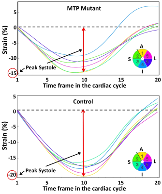 |
9 | Confirming conduction defect in fatty acid oxidation compromised heart using MRI strain imaging
El-Sayed H Ibrahim1, Jason Jarzembowski1, and Suresh Kumar1
1Medical College of Wisconsin, Milwaukee, WI, United States
Healthy heart is a metabolically dynamic organ requiring large energy resource, and fatty acid oxidation (FAO) pathway predominantly supports this energy demands. Disorders of FAO pathways could range from mild and manageable to severe and life-threatening. In this study, we use MRI to evaluate the heart function in mitochondrial trifunctional protein (MTP) mutant rats, where clinical symptoms of this disorder overlap with other less severe FAO deficiencies and are often misdiagnosed. The results showed that MRI strain imaging revealed subclinical cardiac dysfunction in the MTP mutant rats with the degree of strain reduction depending on strain direction and regional location.
|
||
4922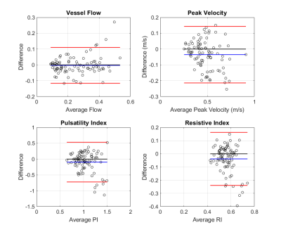 |
10 | Test-Retest Reproducibility of Cerebrovascular Dual-venc 4D Flow MRI
Jackson E. Moore1, Kristina M. Zvolanek1, Maria Aristova1, Ramez Abdalla1, Ann Ragin1, Eric Russell1, Fan Caprio2, Sameer A. Ansari1, Susanne Schnell1,3, and Michael Markl1
1Radiology, Northwestern University, Chicago, IL, United States, 2Neurology, Northwestern University, Chicago, IL, United States, 3Institute of Physics, University of Greifswald, Greifswald, Germany
Cerebrovascular dual-venc 4D Flow MRI has utility in evaluating intracranial hemodynamics - benefitting from full circle of Willis (CoW) volumetric coverage, time-resolved, three-directional velocity encoding, and velocity dynamic range. Analysis of hemodynamic measures was completed using a comprehensive, semi-automated vessel identification and segmentation workflow for 10 subjects who underwent two scans within 30 days. Test-retest analysis of cerebrovascular dual-venc 4D Flow MRI and measures of CoW 3D flow dynamics demonstrated excellent repeatability for flow and peak velocity measures and lower reproducibility for pulsatility and resistive indices. Future work may include a larger cohort or more advanced analysis of velocity-derived parameters.
|
||
The International Society for Magnetic Resonance in Medicine is accredited by the Accreditation Council for Continuing Medical Education to provide continuing medical education for physicians.