Online Gather.town Pitches
Molecular Imaging III
Joint Annual Meeting ISMRM-ESMRMB & ISMRT 31st Annual Meeting • 07-12 May 2022 • London, UK

| Booth # | ||||
|---|---|---|---|---|
4415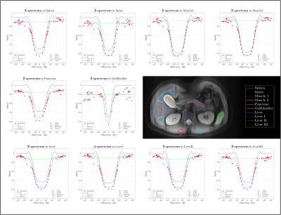 |
1 | CEST and ZAP Measurements of the Abdominal Organs
Vadim Malis1, Jirach Kungsamutr2, Won Bae1, Xiaowei Zhang1, Yoshimori Kassai3, and Mitsue Miyazaki1
1Radiology, UC San Diego, San Diego, CA, United States, 2Bioengineering, UC San Diego, San Diego, CA, United States, 3Canon Medical Systems Corp., Tochigi, Japan
Chemical exchange saturation transfer (CEST) is a molecular imaging technique for specific exchange of hydroxy (-OH, ~1.0 ppm), amine (-NH2, ~2 ppm), and amide (-NH, ~3.5 ppm), whereas ZAP is overall exchange protons of relatively free exchange protons (T2,f) and restricted protons (T2,r) from a wide frequency range of +/– 100 kHz. Combining CEST and ZAP to measure qualitative values of abdominal organ is aiming to be established.
|
||
4416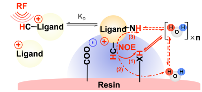 |
2 | Quantification of Electrostatic Molecular Binding Model Using the Water Proton Signal
Chongxue Bie1,2,3, Yang Zhou1,2,4, Peter C. M. van Zijl1,2, Jiadi Xu1,2, Chao Zou4, and Nirbhay N. Yadav1,2
1F.M. Kirby Research Center for Functional Brain Imaging, Kennedy Krieger Institute, Baltimore, MD, United States, 2The Russell H. Morgan Department of Radiology, The Johns Hopkins University School of Medicine, Baltimore, MD, United States, 3Department of Information Science and Technology, Northwest University, Xi'an, China, 4Key Laboratory for Magnetic Resonance and Multimodality Imaging of Guangdong Province, Shenzhen Institute of Advanced Technology, Chinese Academy of Sciences, ShenZhen, China
Saturation transfer MRI has previously been used to probe molecular binding interactions with signal enhancement via the water signal (IMMOBILISE approach). Here, we detailed the relayed nuclear Overhauser effect (rNOE) based mechanisms of this signal enhancement by a four-pool magnetization transfer model, verified the model both using simulations and experimentally (using small charged molecules: arginine, choline, and acetyl-choline). The analytical model can be used to quantify molecular binding affinity, i.e., the dissociation constant (KD). The characterization of the transient binding of small natural substrates paves a pathway towards the detection of receptor-substrate binding in vivo using MRI.
|
||
4417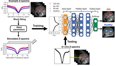 |
3 | Deep learning-based CEST MRI classification of injured kidneys in multiple preclinical models
Chongxue Bie1,2,3, Zheng Han1,2, Peter C. M. van Zijl1,2, Nirbhay N. Yadav1,2, and Guanshu Liu1,2
1F.M. Kirby Research Center for Functional Brain Imaging, Kennedy Krieger Institute, Baltimore, MD, United States, 2The Russell H. Morgan Department of Radiology, The Johns Hopkins University School of Medicine, Baltimore, MD, United States, 3Department of Information Science and Technology, Northwest University, Xi'an, China
Accurate detection of kidney injury is of immense importance for the diagnosis and treatment of acute kidney injury (AKI). While CEST MRI has the potential to reveal the pathophysiological changes on a molecular level, no automatic, CEST-based classification model has been developed. We developed a deep neural network (DNN) to analyze features of the Z-spectral data and to classify injured and healthy renal tissues. The results show that the classification model was capable of reliable prediction of kidney injury among different AKI mouse models. Results correlated well with serum creatinine (SCr) measurement.
|
||
4418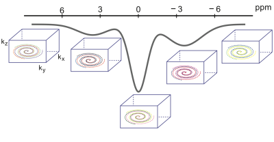 |
4 | CEST MRI Using Golden-Angle Cartesian Acquisition with Sparse Reconstruction
Ding Xia1, Li Feng1, and Xiang Xu1
1Biomedical Engineering and Imaging Institute and Department of Radiology, Icahn School of Medicine at Mount Sinai, New York, NY, United States
In this work, we proposed a novel dynamic CEST MRI framework, which employs golden-angle rotated variable-density Cartesian acquisition with multicoil compressed sensing reconstruction. It enables continuous data acquisition and retrospective reconstruction with desired temporal resolution. We have demonstrated the performance of this new framework for accelerated CEST MRI both in phantom and in human brain at 7T.
|
||
4419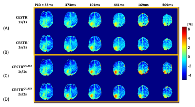 |
5 | Fast Multi-slice Quasi-steady-state (QUASS) APT MRI: Preliminary Results from Brain Tumor Patients at 3 Tesla
Hahnsung Kim1,2, Lisa C. Krishnamurthy 3,4, Kimberly B Hoang5, Ranliang Hu2, and Phillip Zhe Sun1,2
1Yerkes Imaging Center, Yerkes National Primate Research Center, Emory University, Atlanta, GA, United States, 2Department of Radiology and Imaging Sciences, Emory University School of Medicine, Atlanta, GA, United States, 3Center for Visual and Neurocognitive Rehabilitation, Atlanta VA, Decatur, GA, United States, 4Department of Physics & Astronomy, Georgia State University, atlanta, GA, United States, 5Department of Neurosurgery, Emory University School of Medicine, Atlanta, GA, United States
The multi-slice CEST signal evolution was described by the spin-lock relaxation during saturation duration (Ts) and longitudinal relaxation during the relaxation delay time (Td) and post-label delay (PLD), from which the QUASS CEST was generalized to fast multi-slice acquisition. In addition, normal human subjects and tumor patients scans were performed to compare the conventional apparent and QUASS CEST measurements with different Ts, Td, and PLD. Bland-Altman analysis bias of the proposed QUASS CEST effects was much smaller than the PLD-corrected apparent CEST effects (0.03% vs. -0.54%), indicating the proposed fast multi-slice CEST imaging is robust and accurate.
|
||
4420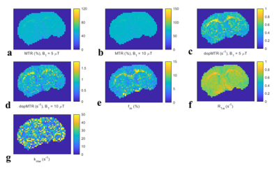 |
6 | Double offsets and powers magnetization transfer ratio (dopMTR) for specific MT MR imaging
zhongliang zu1
1Vanderbilt University Medical Center, Nashville, TN, United States
Magnetization transfer (MT) effect has been analyzed by the MT ratio (MTR) or quantitative MT (qMT). However, because the MTR lacks sensitivity and specificity to MT effect and qMT needs a long scan time, their application into clinical scenarios has been limited. In this paper, we introduce the dopMTR, a new MT data analysis and acquisition method that uses double saturation pulse offsets and powers. Simulations and experiments show that the dopMTR can provide more specific and sensitive quantification of the MT effect than the conventional MTR, with relatively brief acquisition times and easy data processing.
|
||
4421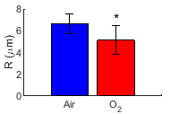 |
7 | Spin-lock dispersion imaging detects changes in the intrinsic susceptibility microstructure of brain induced by oxygen
zhongliang zu1, Fatemeh Adelnia1, Kevin D Harkins1, Feng Wang1, and John C Gore1
1Vanderbilt University Medical Center, Nashville, TN, United States
Previous research has shown that spin-lock imaging with low locking amplitude can quantify the spatial characteristics of intrinsic susceptibility gradients in tissues which likely reflect vascular microstructure. We evaluated the capability of spin-lock dispersion imaging with weak locking fields to detect vasoconstriction in rat brains under oxygen challenges.
|
||
4422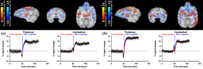 |
8 | Investigation of neurovascular coupling to cannabinoid 1 receptor occupancy: a simultaneous PET/fMRI study on non-human primates
Chi-Hyeon Yoo1, Nisha Rani1, Frederick Bagdasarian1, Sarah Reid1, Changning Wang1, and Hsiao-Ying Wey1
1Athinoula A. Martinos Center for Biomedical Imaging, Department of Radiology, Massachusetts General Hospital, Harvard Medical School, Charlestown, MA, United States
The goal of this study was to investigate relationship between hemodynamic responses and underlying neurochemistry targeting cannabinoid receptor 1 (CB1R) in response to the injection of rimonabant in non-human primates using simultaneous positron emission tomography (PET) and function magnetic resonance imaging (fMRI). Following the rimonabant injection, fast and slow hemodynamic responses were identified throughout the brain. The CB1R occupancy estimates suggested that the radiotracer binding to CB1R was blocked by rimonabant highly in cerebellum and several brain regions. Future PET/fMRI studies with other CB1R-agonists/antagonists and more accurate occupancy estimates will provide further understanding about neurovascular coupling to CB1R.
|
||
4423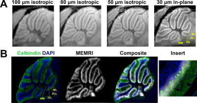 |
9 | Assignment of signal to cell type in MEMRI of the cerebellum
Harikrishna Rallapalli1,2, N. Sumru Bayin3, Hannah Goldman2, Brian J Nieman4, Alan P Koretsky1, Alexandra L Joyner3, and Daniel H Turnbull2
1NINDS, NIH, Bethesda, MD, United States, 2Skirball Institute of Biomolecular Medicine, New York University School of Medicine, New York, NY, United States, 3Developmental Biology, Sloan Kettering Institute, New York, NY, United States, 4Research Operations, The Hospital for Sick Children, Toronto, ON, Canada
Manganese enhanced MRI (MEMRI) has been used to generate layer-specific contrast in preclinical neuroimaging studies, especially in the cerebellum. However, the cell types that contribute most to MEMRI signal have not yet been identified. In this study, we registered high resolution MEMRI to immunohistochemistry of the cerebellum to verify layer localization of signal. We found that the Purkinje cell layer (PCL) was the source of hyperintensity. Next, we manipulated cell types of the PCL using genetically engineered mouse models and quantified MEMRI signal changes. These results strongly suggest Purkinje cells are the primary cellular source of hyperintensity in cerebellar MEMRI.
|
||
4424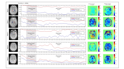 |
10 | Dynamic MTRasym in GlucoCEST MRI using modified DGE
Gustavo Kaneblai1, Maria Concepcion Garcia Otaduy2, Regis Otaviano Franca3, Frederico Perego Costa4, Eduardo Figueiredo5, Thomas Doring6, Marcelo Blois5, Claudia da Costa Leite7, Giovanni Guido Cerri7, and Mitsuharu Miyoshi8
1Digital Innovation, GE Healthcare, Sao Paulo, Brazil, 2Univesidade de Sao Paulo, Sao Paulo, Brazil, 3Hospital Sirio-Libanes, SAO PAULO, Brazil, 4Hosputal Sirio-Libanes, SAO PAULO, Brazil, 5GE Healthcare, SAO PAULO, Brazil, 6GE Healthcare, RIO DE JANEIRO, Brazil, 7Universidade de Sao Paulo, SAO PAULO, Brazil, 8GE Healthcare, Tokyo, Japan
A new method to create B0 corrected dynamic MTRasym glucoCEST maps during glucose administration and uptake without the need of acquiring a full Z-Spectrum with small changes in DGE. Results observed in 3T.
|
||
4425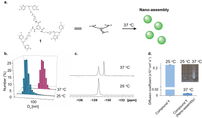 |
11 | In-tissue nano-assembly of Zn2+-responsive agent provides prolonged imaging time-window for longitudinal in vivo MRI studies
Deva Nishanth Tirukoti1, Liat Avram-Biton2, and Amnon Bar-Shir1
1Molecular Chemistry and Material Science, Weizmann Institute of Science, Rehovot, Israel, 2Chemical Research Support, Weizmann institute of Science, Rehovot, Israel
The fast clearance of small molecular probes limits their applicability as responsive agent and prevents their use in longitudinal study. We show here the design and implementation of a tripod-type responsive agent for Zn2+ mapping that assembled to 100nm nanostructure at physiological temperature. The designed fluorinated probe showed capabilities to monitor Zn2+ when combining CEST MRI and 19F-MRI. Upon its intracranial injection the synthetic probe assembled to large entities in the tissue that prevent its clearance from the injection site. This property allows to map its 19F-signal for more than 20 hours and will enable longitudinal studies in the future.
|
||
4426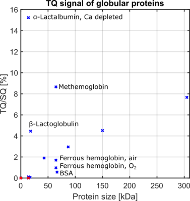 |
12 | Correlation of the 23Na Triple-Quantum Signal with Protein Size
Simon Reichert1, Dennis Kleimaier1, and Lothar Schad1
1Computer Assisted Clinical Medicine, Heidelberg University, Mannheim, Germany
This study demonstrates a correlation of protein size with sodium triple-quantum (TQ) signal. The TQ/SQ ratio of several globular proteins increased with protein size. Furthermore, strong sodium binding had a substantial impact on the TQ signal. The small protein α-Lactalbumin yielded a strong TQ signal due to strong sodium binding to a calcium binding site using a calcium-depleted protein.
|
||
4427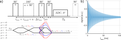 |
13 | Simulation of Spin-3/2 Quadrupole Nuclei Dynamics in Biological Environments for Arbitrary Pulse Sequences
Simon Reichert1, Dennis Kleimaier1, and Lothar Schad1
1Computer Assisted Clinical Medicine, Heidelberg University, Mannheim, Germany
This work presents an open source simulation framework of spin-3/2 NMR dynamics for isotropic and anisotropic environments, and hard pulses. The flexible, modular structure provides a computationally efficient framework to investigate in detail MR signal characteristics using arbitrary pulse sequences. Comparison of simulated and measured data showed a good agreement for the TQTPPI sequence using 23Na. The simulation framework is available as open source on GitHub (https://github.com/SmnReichert/PhaseCycleSim).
|
||
4428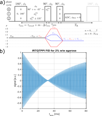 |
14 | Comparison of Double Quantum (DQ) Suppression Methods for Inversion Recovery TQTPPI (IRTQTPPI) Sequence
Simon Reichert1, Dennis Kleimaier1, and Lothar Schad1
1Computer Assisted Clinical Medicine, Heidelberg University, Mannheim, Germany
This study proposes an inversion recovery TQTPPI pulse sequence to create triple quantum (TQ) coherences utilizing the $$$T_1$$$ relaxation pathway. Different double quantum (DQ) suppression methods provide stable fit results with reasonable SNR. The $$$T_1$$$-TQ signal is sensitive to faster motion than the conventional $$$T_2$$$-TQ pathway and thus provides additional information about sodium-protein interactions.
|
||
The International Society for Magnetic Resonance in Medicine is accredited by the Accreditation Council for Continuing Medical Education to provide continuing medical education for physicians.