Oral Session
Deep Learning Image Reconstruction
Joint Annual Meeting ISMRM-ESMRMB & ISMRT 31st Annual Meeting • 07-12 May 2022 • London, UK

| 14:30 | 0498 |
Characterization of cardiac noise in brain quantitative relaxometry MRI data
Quentin Raynaud1, Jérôme Yerly2,3, Ruud B. van Heeswijk2, and Antoine Lutti1
1Clinical Neuroscience, Laboratory for Research in Neuroimaging, Lausanne University Hospital and University of Lausanne, Lausanne, Switzerland, 2Diagnostic and Interventional Radiology, Lausanne University Hospital and University of Lausanne, Lausanne, Switzerland, 3Center for Biomedical Imaging, Lausanne, Switzerland Cardiac-related noise reduces the sensitivity of relaxometry data to brain microstructure properties such as iron or myelin concentration. To address this effect, we aim to characterize cardiac pulsation effects in in-vivo data. We combined a Cartesian pseudo-spiral sampling trajectory with compressed sensing image reconstruction using a temporal total variation regularization to obtain 3D multi-echo images at 12 points of the cardiac cycle. We show that 76% of the full k-space is required in each cardiac bin to maintain 90% of voxels above a 60% sensitivity to physiological noise in the reconstructed images. |
|
14:42 |
0499 |
NLINV-Net: Self-Supervised End-2-End Learning for Reconstructing Undersampled Radial Cardiac Real-Time Data
Moritz Blumenthal1, Guanxiong Luo1, Martin Schilling1, Markus Haltmeier2, and Martin Uecker1,3,4
1University Medical Center Göttingen, Göttingen, Germany, 2Department of Mathematics, University of Innsbruck, Innsbruck, Austria, 3Institute of Medical Engineering, Graz University of Technology, Graz, Austria, 4DZHK (German Centre for Cardiovascular Research), Göttingen, Germany
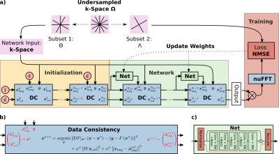
In this work, we propose NLINV-Net, a neural network architecture for jointly estimating the image and coil sensitivity maps of radial cardiac real-time data. NLINV-Net is inspired by NLINV and solves the non-linear formulation of the SENSE inverse problem by unrolling the iteratively regularized Gauss-Newton method, which is improved by adding neural network based regularization terms. NLINV-Net is trained in a self-supervised fashion, which is crucial for cardiac real-time data which lack any ground truth reference. NLINV-Net significantly reduces noise and streaking artifacts compared to reconstructions using plain NLINV.
|
|
| 14:54 | 0500 |
Estimating Noise Propagation of Neural Network Based Image Reconstructio Using Automated Differentiation
Xiaoke Wang1, Danial Ludwig2, Michael Rawson2, Radu V Balan2, and Thomas Ernst1
1Diagnostic Radiology, University of Maryland-Baltimore, Baltimore, MD, United States, 2Mathematics, University of Maryland-College Park, College Park, MD, United States
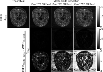
Image reconstructions involving neural networks (NNs) are generally non-iterative and computationally efficient. However, without analytical expression describing the reconstruction process, the compuation of noise propagation becomes difficult. Automated differentiation allows rapid computation of derivatives without an analytical expression. In this work, the feasibility of computing noise propagation with automated differentiation was investigated. The noise propagation of image reconstruction by End-to-end variational-neural-network was estimated using automated differentiation and compared with Monte-Carlo simulation. The root-mean-square error (RMSE) map showed great agreement between automated differentiation and Monte-Carlo simulation over a wide range of SNRs.
|
|
| 15:18 | 0501 |
EigenMRI: Data-driven shallow-learning reconstruction of undersampled MRI data
Ricardo Otazo1,2, Ramin Jafari1, and Masoud Zarepisheh1
1Department of Medical Physics, Memorial Sloan Kettering Cancer Center, New York, NY, United States, 2Department of Radiology, Memorial Sloan Kettering Cancer Center, New York, NY, United States
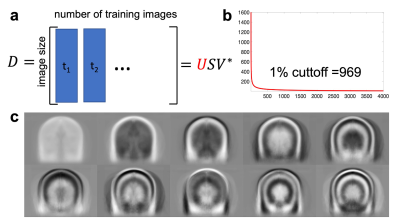
Machine learning, and specifically deep learning, has recently demonstrated high-performance reconstruction of undersampled k-space data. However, training of deep learning reconstruction methods does not take advantage of the thousands of previously acquired images that are stored in clinical databases. Inspired by the eigenfaces method from computer vision, where an image model derived from thousands of pre-existing images is used to identify new faces, this work presents an image reconstruction method named EigenMRI that learns a data-driven regularization approach using thousands of images extracted from a clinical database to reconstruct 3D brain images with 2D acceleration
|
|
| 15:30 | 0502 |
MR Image Reconstruction via Zero-Shot Learned Generative Adversarial Transformers
Salman Ul Hassan Dar1,2, Yilmaz Korkmaz1,2, and Tolga Cukur1,2,3
1Department of Electrical and Electronics Engineering, Bilkent University, Ankara, Turkey, 2National Magnetic Resonance Research Center (UMRAM), Bilkent University, Ankara, Turkey, 3Neuroscience Program, Aysel Sabuncu Brain Research Center, Bilkent University, Ankara, Turkey
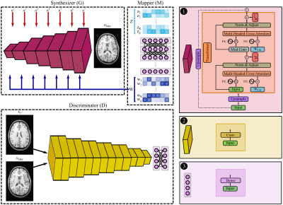
Deep neural network models have demonstrated state-of-the-art performance in MR image reconstruction. These models require information regarding imaging operators during training, which limits their generalization. A recent framework is based on zero-shot learned generative models that learn MR image priors during training and couple imaging operator during inference on test acquisitions. Such models are however based on convolutional architectures that suffer from sub-optimal capture of long range dependencies. Here, we propose a novel architecture based on zero-shot learned generative adversarial transformers that enables efficient capture of long range dependencies via cross-attention transformers while removing reliance on imaging operator during training.
|
The International Society for Magnetic Resonance in Medicine is accredited by the Accreditation Council for Continuing Medical Education to provide continuing medical education for physicians.