Oral Session
Hyperpolarized Metabolic Imaging
Joint Annual Meeting ISMRM-ESMRMB & ISMRT 31st Annual Meeting • 07-12 May 2022 • London, UK

16:45 |
0570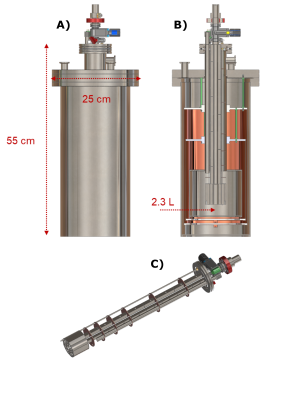 |
A 320 km Hyperpolarization Journey: performing [U-13C, d7]glucose DNP in Copenhagen and Hyperpolarized 13C-MR in Aarhus
Andrea Capozzi1,2, Magnus Karlsson2, Esben Søvsø Szocska Hansen3, Juan Diego Sanchez2, Christoffer Laustsen3, Mathilde Hauge Lerche2, Jacques Van Der Klink1, and Jan Henrik Ardenkjær-Larsen2
1Physics, EPFL, Lausanne, Switzerland, 2Technical University of Denmark, Kgs. Lyngby, Denmark, 3Clinical Medicine, Aarhus University Hospital, Aarhus, Denmark
Dissolution DNP polarizers are expensive, technically demanding and require trained personnel. At the same time, hyperpolarized MR tracers have a half-life short enough to need the polarizer to be placed as close as possible to the MRI scanner. Similarly to PET tracers, being able to receive on demand hyperpolarized contrast agents from a centralized facility would solve this major shortcoming and boost the diffusion of hyperpolarized MR on a wider scale. In this work, using UV-induced non-persistent radicals and purpose build hardware, we demonstrate the first “across-cities-dDNP” experiment.
|
|
16:57 |
0571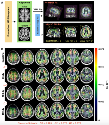 |
Methodological advances in serial hyperpolarized carbon-13 (HP-13C) metabolic imaging of patients with glioma
Adam W Autry1, Sana Vaziri1, Jeremy W Gordon1, Marisa LaFontaine1, Hsin-Yu Chen1, Yaewon Kim1, Javier Villanueva-Meyer1, Susan M Chang2, Jennifer Clarke2,3, Nancy A Oberheim-Bush2,3, Duan Xu1, Janine M Lupo1, Peder EZ Larson1, Daniel B Vigneron1,4, and Yan Li1
1Department of Radiology and Biomedical Imaging, University of California San Francisco, San Francisco, CA, United States, 2Department of Neurological Surgery, University of California San Francisco, San Francisco, CA, United States, 3Department of Neurology, University of California San Francisco, San Francisco, CA, United States, 4Department of Bioengineering and Therapeutic Sciences, University of California San Francisco, San Francisco, CA, United States
This study aimed to improve hyperpolarized carbon-13 (HP-13C) metabolic imaging methodologies for serial monitoring of patients with malignant brain tumors. Several analyses were pursued among 29 patients diagnosed with gliomas and 7 volunteers. Implementation of atlas-based prescription of echo-planar imaging (EPI) demonstrated consistent volumetric coverage (dice:0.967-0.978) and improved hemispheric symmetry in modeled kinetics(p=0.006), which helped highlight tumor. A 2s versus 5s acquisition delay post injection of tracer increased the capture of [1-13C]pyruvate inflow dynamics(p=0.04), while reducing simulated kinetic modeling error. Post-acquisition FID-CSI spectra demonstrated improved referencing of [1-13C]pyruvate f0 using water versus a coil-embedded urea phantom(p=0.00003), thereby maximizing EPI excitation.
|
|
| 17:09 | 0572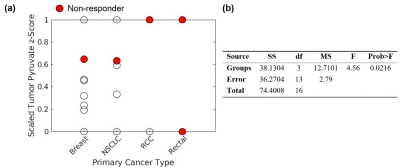 |
Evaluating hyperpolarized [1-13C]pyruvate uptake for predicting response to stereotactic radiosurgery in brain metastases
Nicole I.C. Cappelletto1, Hany Soliman2, Casey Y. Lee1, Nadia D. Bragagnolo1,3, Arjun Sahgal2, Albert P. Chen4, Ruby Endre3, William J. Perks5, Nathan Ma5, Jay S. Detsky2, Chris Heyn6, and Charles H. Cunningham1,3
1Medical Biophysics, University of Toronto, Toronto, ON, Canada, 2Radiation Oncology, Sunnybrook Health Sciences Centre, Toronto, ON, Canada, 3Physical Sciences, Sunnybrook Research Institute, Toronto, ON, Canada, 4GE Healthcare, Toronto, ON, Canada, 5Pharmacy, Sunnybrook Health Sciences Centre, Toronto, ON, Canada, 6Radiology, Sunnybrook Health Sciences Centre, Toronto, ON, Canada
Brain metastases are increasingly being treated with stereotactic radiosurgery; however, 20-30% of treated tumors recur locally post-treatment. Hyperpolarized [1-13C]pyruvate magnetic resonance imaging (HP 13C MRI) is an emerging metabolic imaging modality that measures key metabolic phenotypes indicative of tumor biology. Here we investigate pre-treatment [1-13C]pyruvate uptake – a potential marker of monocarboxylate transporter 1 expression and tumor vascularity – via HP 13C MR images as a predictor of local recurrence. [1-13C]pyruvate uptake establishes a robust predictive model (AUC = 0.73) and, as a result, can inform treatment decisions should the model predict a non-response to SRS.
|
|
| 17:21 | 0573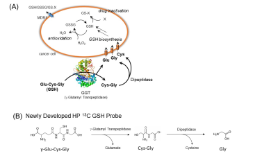 |
Family of Novel Hyperpolarized 13C Molecular Imaging Probes Monitors Real-Time Oxidative Stress in Cancer Metabolism
Kazutoshi Yamamoto1, Yohei Kondo2, Ana Opina3, Deepak Sail3, Natarajan Raju3, Burchelle Blackman3, Ronja M. Malinowski4, Tomohiro Seki1, Shun Kishimoto1, Nobu Oshima1, Yasunori Otowa1, Kota Yamashita1, Daniel R. Crooks1, Jeffrey R. Brender1, Hiroshi Nonaka2, Yutaro Saito2, Peter L. Choyke1, Jan H. Ardenkjær-Larsen 4, James B. Mitchell1, W. Marston Linehan1,
Rolf Swenson3, Shinsuke Sando2, and Murali C. Krishna1
1National Cancer Institute, National Institutes of Health, Bethesda, MD, United States, 2Department of Chemistry and Biotechnology, Graduate School of Engineering, The University of Tokyo, Bunkyo-ku, Japan, 3National Heart, Lung, and Blood Institute, National Institutes of Health, Rockville, MD, United States, 4Department of Health Technology, Technical University of Denmark, Lyngby, Denmark
Molecular imaging can personalize cancer therapies by monitoring treatment responses, where hyperpolarized MRI is becoming an increasingly critical methodology. However, the limited number of available probes, which can interrogate key physiology/biochemistry related to targeted diseases, remains one of the major challenges in this growing field. In this presentation, we successfully demonstrate, for the first time, the advantages of newly synthesized 13C-Glutathione and 13C-N-acetyl cysteine probe in vivo redox status in a complementary manner. This approach serves as powerful non-invasive biomarkers to assess oxidative stress, which plays a vital role in various diseases and can be translatable to the clinical diagnosis.
|
|
| 17:33 | 0574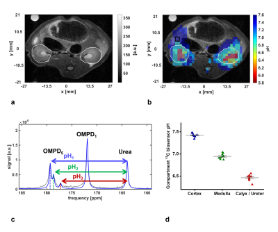 |
In vivo pH imaging in rats using [1,5-13C2]Z-OMPD
Pascal Wodtke1, Martin Grashei1, Frits H. A. van Heijster1, Sandra Sühnel1, Nadine Setzer1, and Franz Schilling1
1Department of Nuclear Medicine, Technical University of Munich, Munich, Germany
We introduce hyperpolarized [1,5-13C2]Z-OMPD as a novel in vivo pH sensor. Upon synthesis from ethyl pyruvate, the molecule appears to be non-toxic and shows good hyperpolarization properties, as indicated by high polarization levels and long T1 relaxation times for both 13C-labels. 13C-chemical shifts show good pH sensitivity in the physiological range, which enables pH imaging in vitro of human blood phantoms of different pH. In vivo pH imaging upon injection in rats reveals previously reported pH compartments in kidneys. It is further demonstrated, that the OMPD1 peak can be used as internal reference, further facilitating the process.
|
|
| 17:45 | 0575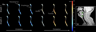 |
Measuring Airflow Velocity in the Upper Airways with 129Xe Phase Contrast MRI: Feasibility in Pediatric Patients with Obstructive Sleep Apnea
Neil James Stewart1,2, Qiwei Xiao2,3, Matthew Willmering2,3, Keith McConnell3, Hui Wang4, Robert Fleck5, Jason Woods2,3, Raouf Amin3, and Alister Bates2,3,5,6
1POLARIS, Imaging Sciences, Department of Infection, Immunity & Cardiovascular Disease, The University of Sheffield, Sheffield, United Kingdom, 2Center for Pulmonary Imaging Research, Cincinnati Children’s Hospital Medical Center, Cincinnati, OH, United States, 3Division of Pulmonary Medicine, Cincinnati Children’s Hospital Medical Center, Cincinnati, OH, United States, 4MR Clinical Science, Philips, Cincinnati, OH, United States, 5Department of Radiology, Cincinnati Children’s Hospital Medical Center, Cincinnati, OH, United States, 6Department of Pediatrics, College of Medicine, University of Cincinnati, Cincinnati, OH, United States We report preliminary HP 129Xe phase contrast (PC) MRI data in pediatric patients with obstructive sleep apnea; a common sleep-related breathing disorder caused by upper airway collapse. The feasibility and safety of 129Xe gas delivery through the patient’s anesthesia mask during tidal breathing under sedation (approximately sleep stage-2) was demonstrated. Dynamic spiral-based PC MRI (temporal resolution 0.5—1s) captured inspiratory and expiratory airflow, with sufficient sensitivity to identify flow features such as high-speed jets corresponding to regions of airway narrowing/obstruction on 1H anatomical MRI. With further data, our work may allow the development of optimized personalized treatment plans. |
|
| 17:57 | 0576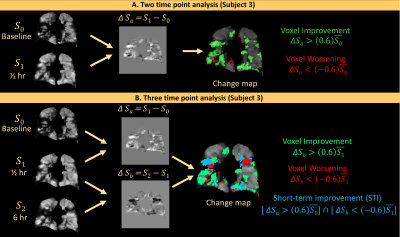 |
Quantifying regional pulmonary ventilation changes in hyperpolarized 3He MRI of asthma subjects following bronchodilator at three timepoints
Ummul AFIA Shammi1, Lucia Flors Blasco2, Talissa Altes3, John P. Mugler4, Craig Meyer4, Jaime Mata5, Kun Qing5, Wilson Miller5, and Robert Thomen1
1Biomedical, Biological & Chemical Engineering, University of Missouri, Columbia, Columbia, MO, United States, 2Keck School of Medicine, University of Southern California, Los Angeles, CA, United States, 3Department of Radiology, University of Missouri, Columbia, Columbia, MO, United States, 4Biomedical Engineering, University of Virginia, Charlottesville, VA, United States, 5Radiology and Medical Imaging, University of Virginia, Charlottesville, VA, United States
Quantification of reginal pulmonary ventilation changes in a longitudinal study with hyperpolarized gas MRI would be beneficial for clinicians evaluating treatment responses. While VDP is a sensitive measure of global lung ventilation, regional ventilation information inherent in imaging is lost. In this work, we present a method of performing voxel-wise comparisons in 3He MRI and producing regional ventilation change maps in asthma patients following bronchodilation with either short-acting or long-acting beta agonists at three timepoints. Regional change maps were congruous with visual assessment of regional ventilation changes and showed the temporal effects of bronchodilator action in patients with severe asthma.
|
The International Society for Magnetic Resonance in Medicine is accredited by the Accreditation Council for Continuing Medical Education to provide continuing medical education for physicians.