Oral Session
Low-Field MRI: New Methods
Joint Annual Meeting ISMRM-ESMRMB & ISMRT 31st Annual Meeting • 07-12 May 2022 • London, UK

| 14:30 | 0313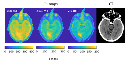 |
Field-Cycling imaging identifies ischaemic stroke at magnetic field strength below 200 mT
Vasiliki Mallikourti1, James Ross1, Oliver Maier2, Markus Bödenler3, Rudolf Stollberger 4, Lionel Broche1, and Mary-Joan MacLeod5
1Aberdeen Biomedical Imaging Centre, University of Aberdeen, Aberdeen, United Kingdom, 2Institute of Medical Engineering, Graz University of Technology, Graz, Austria, 3Institute of eHealth, University of Applied Sciences FH JOANNEUM, Graz, Austria, 4BioTechMed-Graz, Graz, Austria, 5Institute of Medical Sciences, University of Aberdeen, Aberdeen, United Kingdom
Field-Cycling imaging (FCI) is a new imaging technique able to image over a range of low magnetic field levels by rapid switching, to explore the variations in T1 relaxation time as a function of the magnetic field strength, known as T1 dispersion. Here we measured the T1 dispersion contrast from patients with ischaemic stroke. The infarct region in T1 maps corresponded to clinical images. T1 dispersions had different profiles between stroke areas and healthy brain areas, showing that ischaemic strokes can be imaged below 200 mT without contrast agents.
|
|
| 14:42 | 0314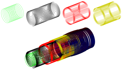 |
An Adaptive Target Field Framework for Complete Low Field MRI System Design
Bart de Vos1, Rob F. Remis2, and Andrew G. Webb1,2
1Leiden University Medical Center, C.J. Gorter Center for High Field MRI, Leiden, Netherlands,, Leiden, Netherlands, 2Delft University of Technology, Circuits and Systems, Delft, Netherlands, Delft, Netherlands
In this work a low field point-of-care system design framework is created using target field methods for all of the hardware components. A new target field method for Halbach-based magnet optimization with variable ring diameter and spacing is derived. Magnet, gradient and RF are combined into a single framework which includes a feedback loop for dealing with the component interdependencies. The result is a pipeline which with a few user inputs can create an optimal magnet, gradient and RF design in minutes.
|
|
| 14:54 | 0315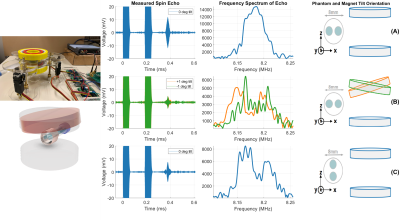 |
2D Imaging without Gradient Coils in a Low-Field Spokes-and-Hub Permanent Magnet
Irene Kuang1, Jason Stockmann2,3, Elfar Adalsteinsson1,4, and Jacob White1
1Electrical Engineering and Computer Science, Massachusetts Institute of Technology, Cambridge, MA, United States, 2A. A. Martinos Center for Biomedical Imaging, Massachusetts General Hospital, Charlestown, MA, United States, 3Harvard Medical School, Boston, MA, United States, 4Institute for Medical Engineering and Science, Massachusetts Institute of Technology, Cambridge, MA, United States
We demonstrate feasibility of 2D imaging without gradient coils in a spokes-and-hub permanent magnet for a low-cost hand-held educational MR scanner. With the ease of physically assembling the imaging system in mind, the magnet was designed asymmetrically, to introduce a built-in z-gradient, and with an axis of rotation for the top magnet, so it can be tilted by either linear or rotational actuators, and generate negative or positive x-gradients. Results from simulation, Hall-effect sensor measurements, and spin echo data show that the permanent-z plus x-gradient can be used to generate orthogonal fields, and disambiguate signals from a two-tube phantom.
|
|
| 15:06 | 0316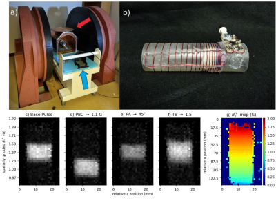 |
Advanced Gradient-Free Selective Excitation Using the Bloch-Siegert Shift
Jonathan B Martin1, Sai Abitha Srinivas1, Christopher Vaughn1, Heng Sun1, Mark Griswold2,3, and William Grissom1,4
1Biomedical Engineering, Vanderbilt University, Nashville, TN, United States, 2Department of Radiology, Case Western Reserve University, Cleveland, OH, United States, 3Biomedical Engineering, Case Western Reserve University, Cleveland, OH, United States, 4Electrical Engineering, Vanderbilt University, Nashville, TN, United States
A class of selective RF pulses which use the Bloch-Siegert shift to localize selective excitation without B0 gradients has recently been proposed. Here we report an improved design method for both the frequency-gradient inducing component pulse and the frequency-selective component pulse, which reduces ripple in the magnetization profile and produces flatter passbands across the excited B1+ range. The pulses’ production of multiphoton resonances across B1+ and their suitability for fast gradient-free CPMG imaging are investigated. They are deployed for the first time on a low-field scanner to produce slice-selective selection.
|
|
| 15:18 | 0317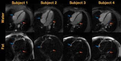 |
Real-Time Water Fat Imaging at 0.55T with Spiral Out-In-Out-In Sampling
Ye Tian1, Yongwan Lim1, and Krishna S. Nayak1
1Ming Hsieh Department of Electrical and Computer Engineering, University of Southern California, Los Angeles, CA, United States
Real-time MRI is a powerful tool to study organ function in a dynamic environment. Many of these regions of interest have both water and fat present, such as the heart and joints. Water/fat separation usually requires multi-echo sequences that utilize chemical shift to distinguish water and fat signals. At 0.55T, the chemical shifts are smaller, making it possible to use long readouts (e.g. spirals) to more efficiently acquire images. Here, we demonstrate spiral out-in-out-in bSSFP imaging combined with region-growing field map estimation for water/fat separation. We demonstrate high quality water/fat separated real-time movies of cardiac function and wrist motion.
|
|
| 15:30 | 0318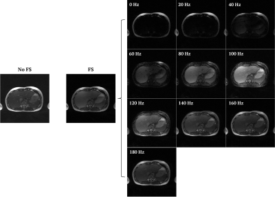 |
Fat-Separated Radial 3D bSSFP Imaging at Low Field Using Frequency-Sweep RF Saturation and XD-GRASP Reconstruction
Ruoxun Zi1, Hersh Chandarana1, and Kai Tobias Block1
1Center for Biomedical Imaging, Department of Radiology, NYU School of Medicine, New York, NY, United States
Low-field systems have recently gained interest because of the potential to make MRI more accessible, but the field reduction comes at expense of low SNR and challenges with fat suppression. This work describes a new concept for fat separation at low field, based on radial acquisition during frequency-sweep RF saturation and combined with frequency-resolved XD-GRASP reconstruction. The approach is demonstrated for free-breathing abdominal imaging using a radial stack-of-stars 3D bSSFP sequence at 0.55T, showing that fat and water can be reliably distinguished based on the response to saturation at different frequencies. Fat-suppressed composite images can be calculated as final step.
|
|
15:42 |
0319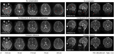 |
Maxwell Field Compensation for 2D Spiral-Ring Turbo Spin-Echo Imaging at 0.55T and 1.5T
Zhixing Wang1, Xue Feng1, Rajiv Ramasawmy2, Adrienne E. Campbell-Washburn2, John P. Mugler III3, and Craig H. Meyer1,3
1Biomedical Engineering, University of Virginia, Charlottesville, VA, United States, 2Cardiovascular Branch, Division of Intramural Research, National Heart, Lung, and Blood Institute, National Institutes of Health, Bethesda, MD, United States, 3Radiology & Medical Imaging, University of Virginia, Charlottesville, VA, United States
Concomitant (Maxwell) fields are problematic for TSE imaging when the readout waveforms vary along the echo train, leading to severe signal dropouts and image blurring. These effects are exaggerated when using prolonged, high-gradient amplitude readouts at lower magnetic-field strengths. The purpose of this work was to implement 2D TSE imaging using annular spiral rings with compensation of self-squared concomitant-gradient terms at the echo time and across echo spacings via gradient waveform modifications, and with compensation of the residual Maxwell- and B0-field induced phase accruals along the readout via image reconstruction. This method reduced artifacts at both 0.55T and 1.5T.
|
 Back to Meeting Home
 Back to the Program-at-a-Glance
The International Society for Magnetic Resonance in Medicine is accredited by the Accreditation Council for Continuing Medical Education to provide continuing medical education for physicians.
 Back to Meeting Home
 Back to the Program-at-a-Glance
 View the Presentation