Digital Poster
MR Fingerprinting & Synthetic MRI
ISMRM & ISMRT Annual Meeting & Exhibition • 03-08 June 2023 • Toronto, ON, Canada

| Computer # | |||
|---|---|---|---|
2352.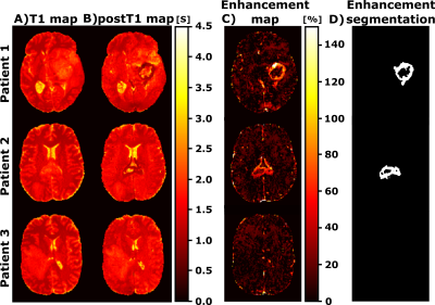 |
1 | Pre and Post contrast Simultaneous Parametric Mapping of Glioblastomas from routine T1 weighted images for Quantitative Enhancement Assessment
Elisa Moya-Sáez1,2, Rodrigo de Luis-García1, Juan A. Hernández-Tamames2, and Carlos Alberola-López1
1University of Valladolid, Valladolid, Spain, 2Erasmus MC, Rotterdam, Netherlands Keywords: MR Fingerprinting/Synthetic MR, Machine Learning/Artificial Intelligence Gadolinium based contrast agents (GBCAs) have the ability to uncover blood brain barrier damage, which appears in the images as contrast enhancement caused by the leakage into the perivascular tissues. However, in clinical practice, this assessment is performed by visual comparison between the weighted images obtained before and after the GBCA injection; enhancement quantification is still an unmet need. In this work we propose a deep learning approach for the computation of pre- and post-contrast parametric maps from conventional T1 weighted images. Results show how those maps can enable an automatic quantification of the tumor enhancement. |
|
2353.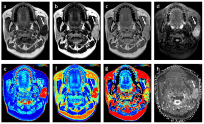 |
2 | Synthetic MRI and FSE-PROPELLER duo diffusion-weighted imaging to differentiate malignant from benign head and neck tumors
Baohong Wen1, Yong Zhang1, Jingliang Cheng1, Tianyong Xu2, Xiaoxia Hua2, and Lizhi Xie2
1the First Affiliated Hospital of Zhengzhou University, Zhengzhou, China, 2GE Healthcare, Shanghai, China Keywords: MR Fingerprinting/Synthetic MR, Head & Neck/ENT Accurate determination of the preoperative classification of histology of head and neck tumor remains a challenge. The aim of the study was to evaluate the feasibility and capability of synthetic MRI and stimulus and fast spin echo diffusion-weighted imaging (FSE-PROPELLER duo-DWI) for the differentiation of malignant from benign head and neck tumors. The results quantitively demonstrated the feasibility and also showed that T2 value is comparable to ADC value, and the combination of T2 and ADC values could provide improved diagnostic efficacy. |
|
2354.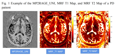 |
3 | Magnetic resonance fingerprinting in patients with Parkinson's disease
Yaping Wu1, Qian Xie2, Ge Zhang1, Yan Bai1, Xipeng Yue1, Wei Wei1, Fangfang Fu1, Nan Meng1, Xianchang Zhang3, Yusong Lin4,5,6, and Meiyun Wang1
1Department of Medical Imaging, Henan Provincial People’s Hospital, Zhengzhou, China, 2School of Computer and Artificial Intelligence, Zhengzhou University, Zhengzhou, China, 3MR Collaboration, Siemens Healthineers Ltd., Beijing, China, 4School of Cyber Science and Engineering, Zhengzhou University, Zhengzhou, China, 5Collaborative Innovation Center for Internet Healthcare, Zhengzhou University, Zhengzhou, China, 6Hanwei IoT Institute, Zhengzhou University, Zhengzhou, China Keywords: MR Fingerprinting/Synthetic MR, MR Fingerprinting, Parkinson's disease This study investigated the utility of 3D high resolution magnetic resonance fingerprinting (MRF) to detect potential brain changes in Parkinson’s disease (PD). MRF T1 and T2 maps of the brains of 22 PD patients and 22 volunteers were non-linearly normalized into the MNI space to explore the group differences using whole-brain analysis. PD patients had significantly higher T1 and T2 values in the right inferior temporal gyrus anterior/posterior division, right temporal fusiform cortex anterior division, right planum polare and red nucleus. Our findings suggest that quantitative parameters acquired from 3D MRF may provide additional information for precise diagnosis of PD. |
|
2355.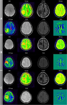 |
4 | Role of MULTIPLEX MRI Study in Evaluating Brain Tumors
Min Gao1, Jun Liu1, Liyun Zheng2, and Yongming Dai3
1Department of Radiology, The Second Xiangya Hospital of Central South University, Changsha, China, 2Shenzhen United Imaging Research Institute of Innovative Medical Equipment, Shenzhen, China, 3MR Collaboration, Central Research Institute, United Imaging Healthcare, Shanghai, China Keywords: MR Fingerprinting/Synthetic MR, MR Fingerprinting Multi-parametric MR imaging methods have been intensively developed and investigated throughout the last decade. The purpose of this study was to utilize the single-scan 3D multi-parametric MRI technique, MULTIPLEX, to characterize and grade intracranial tumors. According to the results, MULTIPLEX MRI can effectively decrease the scan time while obtaining multi-parametric images and mappings. Besides, the simultaneous quantitative estimation of multiple MR parameters can reliably characterize and grade intracranial tumors. |
|
2356.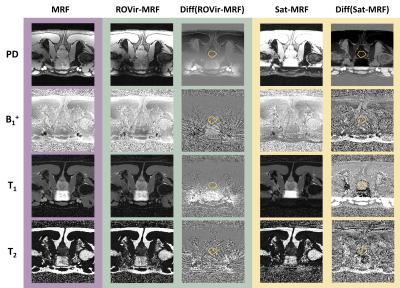 |
5 | Reducing femoral flow artefacts in radial MR Fingerprinting: A comparison of two methods applied to prostate imaging
Kaia Ingerdatter Sørland1, Christopher George Trimble1, Tone Frost Bathen1,2, Mattijs Elschot1,2, and Martijn A. Cloos3
1Department of Circulation and Medical Imaging, Norwegian University of Science and Technology, Trondheim, Norway, 2Department of Radiology and Nuclear Medicine, St. Olavs hospital, Trondheim University Hospital, Trondheim, Norway, 3Centre for Advanced Imaging, The University of Queensland, Brisbane, Australia Keywords: MR Fingerprinting/Synthetic MR, Artifacts High acceleration factors in radial magnetic resonance fingerprinting (MRF) of the prostate lead to strong streaking artefacts from flow in the femoral blood vessels, distorting quantitative T1 and T2 measurements. We here compare two approaches to mitigate these artefacts, namely incorporating regional saturation bands in the MRF sequence (Sat-MRF) or applying region-optimized virtual coils to suppress signal from select spatial regions before image reconstruction (ROVir-MRF). Results from seven asymptomatic volunteers show that both methods efficiently reduce signal in the region of the femoral vessels, but there are differences in retained prostate signal to noise ratio and sensitivity to T1. |
|
2357.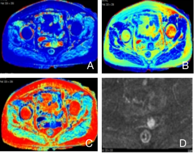 |
6 | Correlation of quantitative synthetic mapping imaging with cervical squamous carcinoma pathology
Qi Zhao1, Zhihua Pan1, Mingli Jin1, Kebin Yu1, Yin Jiang1, and Miaoqi Zhang2
1The 2nd Affiliated Hospital of Chengdu Medical College Nuclear Industry 416 Hospital, Chengdu, China, 2GE Healthcare, Beijing, China Keywords: MR Fingerprinting/Synthetic MR, MR Value Clinical and pathological staging are vital for management of Cervical squamous carcinoma (CSC). In this study, using synthetic MR technique, we investigate quantitative T1, T2 and PD measurements in different clinical and pathological stage groups. Performance of those quantitative measurements in diagnosing poorly-differentiated CSC is also investigated. We find T2 values are higher in late clinical stage CSC. Results also show a correlation between quantitative measurements and the degree of pathological differentiation. ROC analysis demonstrate that T1 and T2 values can differentiate poorly-differentiated CSC from intermediate and well differentiated CSC1. |
|
2358.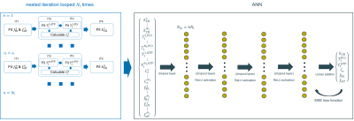 |
7 | A nested iteration artificial neural network approach for efficient high dimensional parameter estimation in 31P-MRF
Mark Widmaier1,2,3, Zirun Wang1, Song-I Lim1,2,3, Daniel Wenz1,2, and Lijing Xin1,2
1CIBM Center for Biomedical Imaging, Lausanne, Switzerland, 2Animal Imaging and Technology, Ecole Polytechnique Federale de Lausanne (EPFL), Lausanne, Switzerland, 3Laboratory of functional and metabolic imaging, Ecole Polytechniqe Federale de Lausanne (EPFL), Lausanne, Switzerland Keywords: MR Fingerprinting/Synthetic MR, Machine Learning/Artificial Intelligence, MRS, Phosporus, Creatine Kinase, MRF A new magnetization transfer 31P Magnetic Resonance Fingerprinting (31P-MRF) technique is emerging to measure the creatine kinase (CK) chemical exchange rate kCK. The inherent obstacle of the exponential growth in the size of dictionaries with the number of free parameters, was overcome by introducing the nested iteration interpolation method (NIIM). To further reduce the processing time and cope with a nonlinear behaviour, we employed an artificial neuron network (ANN), instead of an interpolation method (IPM) as in the original approach. The nested iteration ANN method (NIAN) is compared with NIIM using simulation data and in vivo 31P-MRF data. |
|
2359.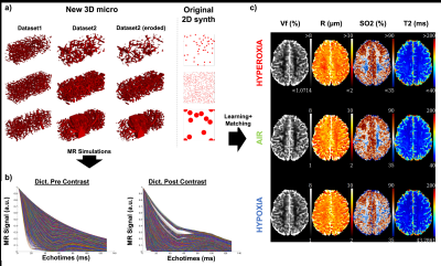 |
8 | MR Vascular Fingerprinting with 3D realistic blood vessel structures and machine learning to assess oxygenation changes in human volunteers
Aurélien Delphin1, Thomas Coudert1, Audrey Fan2, Michael E. Moseley3, Greg Zaharchuk3, and Thomas Christen1
1Univ. Grenoble Alpes, INSERM U1216, Grenoble Institut Neurosciences, GIN, Grenoble, France, 2Biomedical Engineering, University of California Davis, Davis, CA, United States, 3Department of Radiology, Stanford University, Stanford, CA, United States Keywords: MR Fingerprinting/Synthetic MR, Oxygenation The MR vascular fingerprinting (MRvF) approach extends the concept of MR fingerprinting to the study of microvascular properties and functions. Encouraging results have been obtained in healthy human volunteers as well as in stroke and tumor models in rats. However, it has been suggested that the method has a low sensitivity to blood oxygenation measurements. We improved the MRvF approach by using simulations with 3D realistic blood vessels from animal microscopy, new fingerprint-pattern organization and machine learning tools. The method was tested in retrospective data acquired in healthy-human volunteers while breathing different gas mixtures (Hyperoxia (100%O2), Normoxia (21%O2), hypoxia (14%O2)). |
|
2360.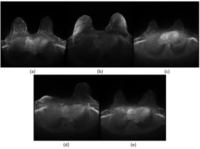 |
9 | Latent Diffusion Models Allow Generation of Synthetic Breast MRI DCE-MIPs
Lukas Folle1, Lorenz Kapsner2,3, Andreas Maier1, Michael Uder2, Sabine Ohlmeyer2, and Sebastian Bickelhaupt2
1Computer Science, Friedrich-Alexander-Universität Erlangen-Nürnberg, Erlangen, Germany, 2Institute of Radiology, Universitätklinikum Erlangen-Nürnberg, Friedrich-Alexander-Universität Erlangen-Nürnberg, Erlangen, Germany, 3Universitätklinikum Erlangen-Nürnberg, Friedrich-Alexander-Universität Erlangen-Nürnberg, Medizinisches Zentrum für Informations- und Kommunikationstechnik, Erlangen, Germany Keywords: MR Fingerprinting/Synthetic MR, Breast The training of neural networks for classification or segmentation of medical images requires large amounts of training data. Sharing of these datasets is commonly difficult due to legislation and privacy constraints of medical data. In this work, we demonstrate the utility of latent diffusion models that allow the generation of synthetic samples of dynamic contrast-enhanced breast MRI-derived maximum intensity projections of subtraction series. Whilst the image quality of the generated data is high as demonstrated by a radiologist evaluation, further steps are envisioned to derive specific compounds of data, e.g., BI-RADS, FGT, or BPE classes. |
|
2361.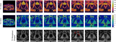 |
10 | 3D High Resolution MR Fingerprinting for prostate cancer
Jesus Ernesto Fajardo1, Anna Lavrova1, Vikas Gulani1, and Yun Jiang1,2
1Department of Radiology, University of Michigan, Ann Arbor, MI, United States, 2Department of Biomedical Engineering, University of Michigan, Ann Arbor, MI, United States Keywords: MR Fingerprinting/Synthetic MR, MR Fingerprinting, prostate cancer We developed a 3D MR Fingerprinting method with B0 correction for imaging the prostate gland. T1 and T2 maps with the spatial resolution of 1 x 1 x 3 mm3 were obtained from phantom and in-vivo experiments, demonstrating the potential of performing accurate and high-resolution tissue quantification of prostate cancer. |
|
2362.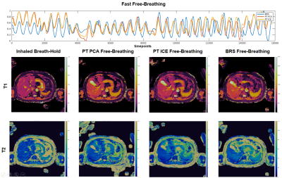 |
11 | Towards Prospective Free-Breathing Abdominal Magnetic Resonance Fingerprinting
Madison Kretzler1, Xinzhou Li2, Leonardo Kayat Bittencourt3,4, Mark Griswold1, and Rasim Boyacioglu1
1Radiology, Case Western Reserve University, Cleveland, OH, United States, 2MR R&D Collaborations, Siemens Medical Solutions USA, Inc., St. Louis, MO, United States, 3Radiology, University Hospitals Cleveland Medical Center, Cleveland, OH, United States, 4School of Medicine, Case Western Reserve University, Cleveland, OH, United States Keywords: MR Fingerprinting/Synthetic MR, Liver, prospective, Free-breathing, Pilot tone A Pilot Tone (PT) based comparison study is presented for abdominal free-breathing MRF using a previously validated PT extraction and vendor developed extraction methods. The initial steps towards prospective processing of the data for immediate availability of the free-breathing MRF quantitative maps on the console are demonstrated. In vivo comparison results for various free-breathing patterns are shown with consistent agreement between respiratory motion in PT extraction methods. Extraction of the respiration signal from PT via the vendor reconstruction software is the first step towards prospective free-breathing abdominal MRF. |
|
2363.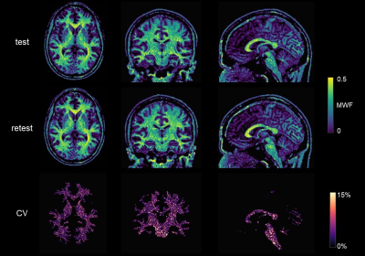 |
12 | Repeatability and reproducibility of MRF-based Myelin Water Fraction maps of healthy human brains
Marta Lancione1, Matteo Cencini1, Guido Buonincontri1, Jan Kurzawski2, Joshua D Kaggie3, Tomasz Matys3, Ferdia A Gallagher3, Graziella Donatelli4,5, Paolo Cecchi4,5, Mirco Cosottini6, Nicola Martini7, Francesca Frijia7, Domenico Montanaro1, Pedro A Gómez4, Rolf F Schulte8, Alessandra Retico9, Laura Biagi1, and Michela Tosetti1
1IRCCS Stella Maris, Pisa, Italy, 2New York University, New York, NY, United States, 3University of Cambridge, Cambridge, United Kingdom, 4IMAGO7 Foundation, Pisa, Italy, 5Azienda Ospedaliero-Universitaria Pisana, Pisa, Italy, 6University of Pisa, Pisa, Italy, 7Fondazione Toscana Gabriele Monasterio, Pisa, Italy, 8GE Healthcare, Munich, Germany, 9Istituto Nazionale di Fisica Nucleare, Pisa, Italy Keywords: MR Fingerprinting/Synthetic MR, MR Fingerprinting, Myelin, Reproducibility Myelin Water Fraction (MWF) can measure White Matter (WM) myelination and integrity and can be quantified using Magnetic Resonance Fingerprinting (MRF) which allows short scan time. In this work, we assessed the repeatability and the reproducibility of MWF using 3D SSFP MRF in a traveling head study performed on healthy volunteers scanned at five different scanners from the same vendor. We computed coefficients-of-variation to estimate voxelwise variability in WM and GLM analysis to measure biases. We reported a variability of ∼3% for repeated scans at the same site and ∼5% for different sites, with an average bias of 5%. |
|
2364.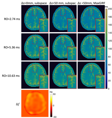 |
13 | Efficient MRF at 0.55T with long readouts and concomitant field effects correction
Zhibo Zhu1, Nam Gyun Lee2, Ye Tian1, and Krishna S. Nayak1
1Department Of Electrical and Computer Engineering, University of Southern California, Los Angeles, CA, United States, 2Department Of Biomedical Engineering, University of Southern California, Los Angeles, CA, United States Keywords: MR Fingerprinting/Synthetic MR, Brain We demonstrate FISP-MRF at 0.55T with improved efficiency using longer spiral readouts and concomitant field effect correction. 2D axial FISP-MRF is performed with 3 different readout durations and at 2 different off-isocenter locations, reconstructed without and with MaxGIRF concomitant field correction. Spatial blurring induced by concomitant field effect was successfully mitigated. In-vivo MRF white matter T2 achieved tighter distribution with longer readouts, e.g., the coefficient of variation decreased from 9.9% to 4.9%. |
|
2365.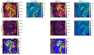 |
14 | Inline calculation of Extracellular Volume Maps using Cardiac Magnetic Resonance Fingerprinting
Alexander Fyrdahl1,2, Jenny Castaings2, Rebecka Steffen Johansson1, Kelvin Chow3, Jannike Nickander1,2, Nicole Seiberlich4, and Jesse I Hamilton4
1Department of Molecular Medicine and Surgery, Karolinska Institutet, Stockholm, Sweden, 2Department of Clinical Physiology, Karolinska University Hospital, Stockholm, Sweden, 3MR R&D Collaborations, Siemens Medical Solutions USA Inc., Chicago, IL, United States, 4Department of Radiology, University of Michigan, Ann Arbor, MI, United States Keywords: MR Fingerprinting/Synthetic MR, Cardiovascular This work describes a pipeline for fully inline generation of T1, T2 and extracellular volume (ECV) maps from cardiac Magnetic Resonance Fingerprinting (MRF) data. Reconstruction of multi-parametric tissue property maps using cardiac MRF have recently been made possible directly on the scanner by replacing the need to perform Bloch equation simulations with a neural network. MRF-derived T1, T2 and ECV values were acquired and compared to MOLLI and T2p-SSFP in 5 successive clinical patients. |
|
2366.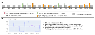 |
15 | Highly efficient T1, T2 and Diffusion-prepared radial Magnetic Resonance Fingerprinting
Carlos Velasco1, Carlos Castillo-Passi1,2, Nadia Chaher1, Alkystis Phinikaridou1, René M. Botnar1,2, and Claudia Prieto1,2
1School of Biomedical Engineering & Imaging Sciences, King's College London, London, United Kingdom, 2Institute for Biological and Medical Engineering, Pontificia Universidad Católica de Chile, Santiago, Chile Keywords: MR Fingerprinting/Synthetic MR, Diffusion/other diffusion imaging techniques In this study we present a golden-angle-radial efficient magnetic resonance fingerprinting approach that enables simultaneous T1, T2 and ADC quantification in a single scan of ~18s and offers the possibility to be extended to a multi-echo acquisition for additional water-fat separation estimation. A quantitative comparison between reference maps and the proposed MRF maps has shown excellent agreement, and a proof-of-concept brain MRF multiparametric T1, T2 and ADC has shown comparable image quality compared to the longer and sequential clinical reference scans. |
|
2367.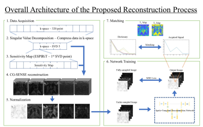 |
16 | Spatio-Temporal Reconstruction Neural Network for 3D MR fingerprinting of the Prostate
Jae-Yoon Kim1, Jae-Hun Lee1, Dongyeob Han2, Moon Hyung Choi3, and Dong-Hyun Kim1
1Department of Electrical and Electronic Engineering, Yonsei Univ., Seoul, Korea, Republic of, 2Siemens Healthineers Ltd, Siemens Korea, Seoul, Korea, Republic of, 3Department of Radiology, Eunpyeong St. Mary's Hospital, College of Medicine,The Catholic University of Korea, Seoul, Korea, Republic of Keywords: MR Fingerprinting/Synthetic MR, Image Reconstruction, Prostate Magnetic Resonance Fingerprinting (MRF) is a technology that computes T1, T2 parameters from time-evaluated signals. However, long scanning time in obtaining fully-sampled data is a challenging point while reducing the sampling rate results in poor reconstructed data quality. Here, we propose a spatio-temporal deep learning network for reconstruction from the under-sampled MRF data. According to the retrospective reconstructed results, the proposed method could produce the T1 and T2 maps of high fidelity similar to the fully-sampled ground-truth. |
|
2368.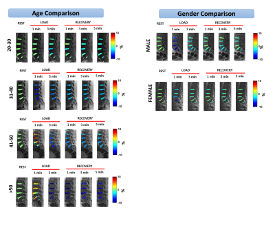 |
17 | Characterization of age- and gender-dependent differences in intervertebral disc strain using 3D-GRASP MRI under static mechanical loading
Rajiv G Menon1, Hector L de Moura1, Richard Kijowski1, and Ravinder R Regatte1
1Radiology, NYU Grossman School of Medicine, New York, NY, United States Keywords: MR Fingerprinting/Synthetic MR, Tissue Characterization, Intervertebral disc The goal of this study was to assess the effect of age and gender in IVD characterization using a multiparameter MR Fingerprinting technique that can quantify T1, T2 and T1rho in a clinically feasible time. Seventeen healthy subjects were recruited. The goal of this study was to assess the effect of age and gender in IVD characterization using a multiparameter MR Fingerprinting technique that can quantify T1, T2 and T1rho in a clinically feasible time. This study suggests that the use of MR fingerprinting is a useful tool to gain insights to pathology resulting in IVD degeneration. |
|
2369.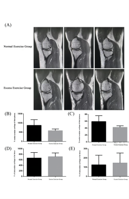 |
18 | Synthetic MRI derived relaxation mapping in evaluating cartilaginous degeneration caused by prolonged excessive exercise
Likai Yu1, Xiaoqing Shi1, Lishi Jie1, Yibao Wei1, Weiqiang Dou2, Shaowei Liu3, and Nongshan Zhang3
1Nanjing University of Chinese Medicine, Nanjing, China, 2GE Healthcare, MR Research China, Beijing, China, 3Jiangsu Province Hospital of Chinese Medicine, Nanjing, China Keywords: Data Analysis, Osteoarthritis The purpose of this study was to investigate whether prolonged overexertion can lead to cartilage degeneration through Synthetic MRI derived relaxation maps. In this study, 30 participants were recruited and measured with T1 and T2 mapping derived by Synthetic MRI. There was a statistically significant trend in cartilage T1 and T2 values in the long-term exercise group compared to the normal exercise group. Based on altered T1 and T2 relaxation properties of cartilage tissues, we conclude that chronic overexercise may lead to cartilage degeneration. |
|
2370.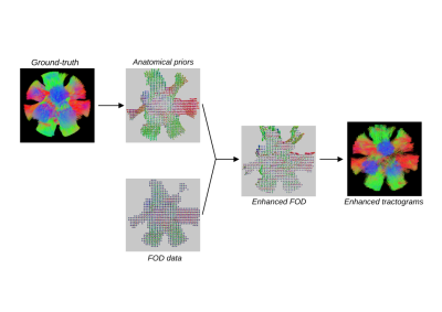 |
19 | Anatomical priors informed tractography. Evaluation on the DiSCo synthetic dataset
Thomas Durantel1, Julie Coloigner1, Emmanuel Caruyer1, Gabriel Girard2,3, and Olivier Commowick1
1Univ Rennes, INRIA, CNRS, INSERM, IRISA UMR 6074, Empenn ERL U-1228, F-35000, Rennes, France, 2Signal Processing Laboratory (LTS5), Ecole Polytechnique Fédérale de Lausanne (EPFL), Lausanne, Switzerland, 3Department of Computer Science, Université de Sherbrooke, Sherbrooke, QC, Canada Keywords: Data Processing, Tractography & Fibre Modelling White matter fiber tracking using diffusion magnetic resonance imaging (DWI) provides a powerful approach to map brain connections, but they are not completely reliable both in academic and clinical context. This is because most methods infer global connectivity from local information,leading to a high rate of false positive. We propose a method to include anatomical prior in the tractography algorithm. We show on synthetic DiSCo data that including this prior on two state-of-the-art tractography algorithms could improve the overall shape of the tractograms. |
|
2371.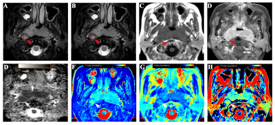 |
20 | Synthetic MRI in differentiating benign from metastatic retropharyngeal lymph nodes: combination with diffusion-weighted imaging
Peng Wang1, Shudong Hu2, Weiqiang Dou3, Jie Shi3, and Heng Zhang1
1Affiliated hospital of Jiangnan university, Wuxi, China, 2ffiliated hospital of Jiangnan university, Wuxi, China, 3GE Healthcare, MR Research China, Beijing, China Keywords: Head & Neck/ENT, Quantitative Imaging This study aimed to evaluate MAGiC imaging (one synthetic MRI technique, syMRI) and its combination with diffusion-weighted imaging (DWI) in discriminating benign from metastatic retropharyngeal lymph nodes (RLNs). With MAGiC derived relaxation parameters, 58 patients with 21 benign and 42 metastatic RLNs were measured. The resultant T1, T2, PD and T1SD values showed significant different values between benign and metastatic RLNs with an optimal diagnostic performance from T1SD. Moreover, the combination of MAGiC, DWI, and morphological features demonstrated a significantly improved performance on overall diagnosis. |
|
The International Society for Magnetic Resonance in Medicine is accredited by the Accreditation Council for Continuing Medical Education to provide continuing medical education for physicians.