Digital Poster
Device & Multimodal I
ISMRM & ISMRT Annual Meeting & Exhibition • 03-08 June 2023 • Toronto, ON, Canada

| Computer # | |||
|---|---|---|---|
4382.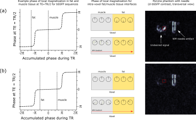 |
121 |
Dephased bSSFP for the Delineation of Interventional Devices
Jonas Frederik Faust1,2,
Peter Speier1,
Axel Joachim Krafft1,
Sunil Patil3,
Mark E. Ladd2,4,
and Florian Maier1
1Siemens Healthcare GmbH, Erlangen, Germany, 2Ruprecht-Karls-Universität Heidelberg, Heidelberg, Germany, 3Siemens Medical Solutions USA Inc., Malvern, PA, United States, 4German Cancer Research Center (DKFZ), Heidelberg, Germany Keywords: Interventional Devices, MR-Guided Interventions Dephased MRI, e.g., used for the delineation of metallic interventional devices, is the reconstruction of images from a shifted k-space and can be achieved by introducing additional “White-Marker” magnetic field gradient moments into the acquisition scheme. A prototype 3D radial sequence was implemented to analyze and compare the artifact of a biopsy needle in a gel phantom for dephased GRE and dephased bSSFP contrast. We found dephased bSSFP to show improved artifact symmetry in comparison with dephased GRE. Undesired signal attributed to fat tissue interfaces in an ex-vivo phantom was successfully suppressed by introducing an altered bSSFP phase cycling. |
|
4383.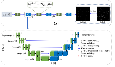 |
122 |
Susceptibility-based Positive Contrast Imaging of MR Compatible
Metallic Devices Using Model-based Deep Learning
Caiyun Shi1,2,
Jing Cheng1,
Xin Liu1,
Hairong Zheng1,
Yanjie Zhu1,
Dong Liang1,3,
and Haifeng Wang1
1Lauterbur Research Center for Biomedical Imaging, Shenzhen Institutes of Advanced Technology, Chinese Academy of Sciences, Shenzhen, China, Shenzhen, China, 2Shenzhen College of Advanced Technology, University of Chinese Academy of Sciences, Shenzhen, China, Shenzhen, China, 3Research Centre for Medical AI, Shenzhen Institutes of Advanced Technology, Chinese Academy of Science, Shenzhen, China, Shenzhen, China Keywords: Interventional Devices, Machine Learning/Artificial Intelligence Susceptibility-based positive contrast MR imaging exhibits excellent efficacy for visualizing the MR compatible metallic devices, by taking advantage of their high magnetic susceptibility. In this work, a model-based deep learning architecture with U-net is developed to realize the 3D susceptibility-based positive contrast MR imaging on real phantom experiments. We train the network on synthetic data to generate positive contrast images from magnetic field maps for localizing the seeds from their surroundings and demonstrate the potential of the deep learning implementation. |
|
4384.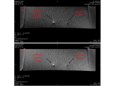 |
123 |
Passive Device Tracking for interventional MRI with Ferric Ion
Chelated Natural Melanin Nanoparticles
Engin Baysoy1,
Gizem Kaleli-Can2,
Alpay Özcan3,
and Chunlei Liu4
1Department of Electrical Engineering and Computer Sciences, UC Berkeley, BERKELEY, CA, United States, 2Department of Biomedical Engineering, İzmir Demokrasi University, İzmir, Turkey, 3Department of Electrical and Electronics Engineering, Boğaziçi University, İstanbul, Turkey, 4Department of Electrical Engineering and Computer Sciences, UC Berkeley, Berkeley, CA, United States Keywords: Interventional Devices, MR-Guided Interventions, Passive Tracking, Melanin Nanoparticles, MRI devices, interventional MRI Natural melanin nanoparticles (MNPs) labeled with paramagnetic ions provide a potential MRI contrast enhancing material alternative to current MRI contrast agents due to their biocompatibility, biodegradability, metal, and biomolecule chelation properties. In this study, we investigated T1 shortening effect of Fe3+ chelated MNPs solution compared to gadolinium-based contrast agents under 3T MRI. Then, we examined traceability of MNP-Fe3+ ions deposited interventional nitinol guidewire in MR scans. Results clearly showed that the coating of Fe3+ chelated MNPs over the surface of MRI compatible instruments will increase traceability of interventional devices in MRI. |
|
4385.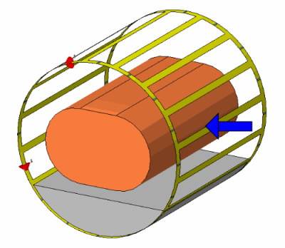 |
124 |
Performance of a rotatable Tx/Rx body coil for MR-guided
particle therapy
Kilian A. Dietrich1,2,3,4,
Sebastian Klüter2,5,6,
Jürgen Debus2,3,4,5,6,7,8,9,10,
Mark E. Ladd1,3,9,
and Tanja Platt1,2,4
1Medical Physics in Radiology, German Cancer Research Center (DKFZ), Heidelberg, Germany, 2Department of Radiation Oncology, Heidelberg University Hospital, Heidelberg, Germany, 3Faculty of Physics, Heidelberg University, Heidelberg, Germany, 4Clinical Cooperation Unit Radiation Oncology, German Cancer Research Center (DKFZ), Heidelberg, Germany, 5National Center for Radiation Research in Oncology (NCRO), Heidelberg, Germany, 6Heidelberg Institute of Radiation Oncology (HIRO), Heidelberg, Germany, 7National Center for Tumor Diseases (NCT), Heidelberg, Germany, 8German Cancer Consortium (DKTK), Heidelberg, Germany, 9Faculty of Medicine, Heidelberg University, Heidelberg, Germany, 10Department of Radiation Oncology, Heidelberg Ion-Beam Therapy Center (HIT), Heidelberg, Germany Keywords: Interventional Devices, MR-Guided Interventions, Particle therapy Electromagnetic field simulations were performed at 1.5T to characterize the imaging capabilities of an RF body coil that is compatible with MR-guided radiotherapy and that can be rotated to achieve a flexible coil to fixed beam orientation. Transmit and receive field characteristics of the RF coil were calculated inside a phantom, and the homogeneity and power efficiency were analyzed for 24 different angles of rotation. |
|
4386.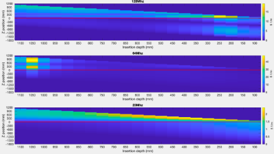 |
125 |
Simulation of the Electric Transfer Function of Partially
Inserted Guidewires
Felipe Godinez1,
Özlem Ipek2,3,
Jeffrey Hand2,3,
Fatima Ben Haj4,
Joseph V Hajnal2,3,
and Shaihan J Malik2,3
1University of California Davis, Sacramento, CA, United States, 2School of Biomedical Engineering and Imaging Sciences, King's College London, London, United Kingdom, 3Centre for the Developing Brain, King's College London, London, United Kingdom, 4Davis Senior High School, Davis, CA, United States Keywords: Interventional Devices, Safety Endovascular interventional devices can heat when used in an MRI system. The heating mechanism can be evaluated using the electric transfer function. In this work we evaluated the TF for a partially inserted guidewire as used in practice, at frequencies corresponding to 0.55T, 1.5T, and 3T. The study revealed that the transfer function varies dramatically with insertion depth for some frequencies and not others. |
|
4387.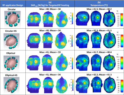 |
126 |
Advanced RF antenna arrangements to improve the efficacy of
Thermal Magnetic Resonance of Glioblastoma Multiforme at 7.0T,
9.4Tand 10.5T
Nandita Saha1,2,
Andre Kuehne3,
Jason M. Millward1,
and Thoralf Niendorf1,4
1Berlin Ultrahigh Field Facility (B.U.F.F.), Max-Delbrück Center for Molecular Medicine in the Helmholtz Association, Berlin, Germany, 2The Charité – Universitätsmedizin, Berlin, Germany, 3MT MedTech Engineering GmbH, Berlin, Germany, 4Experimental and Clinical Research Center (ECRC), a joint cooperation between the Charité Medical Faculty and the Max-Delbrück Center for Molecular Medicine in the Helmholtz Association, Berlin, Germany Keywords: Interventional Devices, MR-Guided Interventions, Thermal MR This work proposes the concept of four RF applicators combining SGBT dipole and loop building block antenna array for diagnostic proton (1H) imaging and thermal intervention at 7.0T, 9.4T and 10.5T. We demonstrated the relationship between spatial arrangement of the antennas in RF applicator and the SAR profile which further related to temperature rise for targeted thermal intervention of deep-seated brain tumors. This approach of design, simulation, optimization of EM power deposition followed by calculation of the resulting temperature distribution can be adapted as a pre-routine to conduct optimization result for individual patient for further treatment planning. |
|
4388.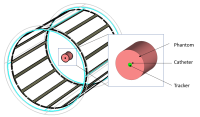 |
127 |
High permittivity material for catheter tracking in
interventional imaging
Yunkun Zhao1 and
Xiaoliang Zhang1
1Biomedical Engineering, State University of New York at Buffalo, Buffalo, NY, United States Keywords: Interventional Devices, MR-Guided Interventions This study presents a technique of using high permittivity dielectric materials for catheter tracking in interventional endovascular imaging procedures. The design includes a high dielectric material cylinder at the tip of the catheter. Given the small size and limited space of the catheters, it is challenging to implement the current catheter tracking techniques. In the proposed approach, the high permittivity material mounted on tip of the catheter can amplify the local magnetic fields and makes the catheter tip visible. This method doesn’t need any feeding cables and is not sensitive to the frequency shift which makes it easy to implement. |
|
4389. |
128 |
Design of a well decoupled 4-channel catheter Radio Frequency
coil array for endovascular MR imaging at 3T
KOMLAN PAYNE1,
Yunkun Zhao1,
Leslie Lei Ying1,
and Xiaoliang Zhang1
1Department of Biomedical Engineering, University at Buffalo, Buffalo, NY, United States Keywords: Interventional Devices, Blood vessels Catheter-based radio frequency (RF) coils for interventional MRI are proposed as a means of obtaining high resolution images with significant advantages over conventional external signal detecting RF coils. A 4-channel catheter array coil was designed to improve the power transmission and SNR using parallel imaging and ultra-sensitive imaging with maximum proximity inside a blood vessel phantom. This design can be scaled to smaller/larger form to fit in tiny/wide vascular structure to provide the required B1 fields and sufficient decoupling among the resonant elements. Numerical simulation was employed to validate the performance and the feasibility of the proposed design. |
|
4390.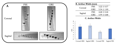 |
129 |
Development and Evaluation of an MR-visible Interventional
Microcatheter
Bridget F Kilbride1,
Teri Moore1,
Alastair J Martin1,
Mark W Wilson1,
and Steven W Hetts1
1Radiology and Biomedical Imaging, University of California, San Francisco, San Francisco, CA, United States Keywords: Interventional Devices, Interventional Devices Intra-arterial chemotherapy following blood-brain barrier opening (BBBO) can be a highly effective therapeutic delivery mechanism when treating primary and metastatic disease in the brain. Osmotic blood-brain barrier disruption has long been performed under x-ray guidance but has yet to gain clinical traction partially due to outcome variability. MRI is advantageous for its superior soft tissue contrast and real-time perfusion territory validation. We propose a passive microcatheter to provide real-time feedback of catheter positioning during BBBO under MRI guidance. In a phantom, we quantitated susceptibility artifacts at 3T. The catheter showed promise as a new MRI-visible tool to enhance BBBO procedures. |
|
4391.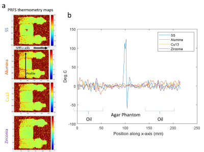 |
130 |
Initial experiences assessing the impact of different materials
for needle-based interventional devices using advanced DWI and
MR thermometry
James H Holmes1,2,3,
Collin J Buelo4,5,
Ruiqi Geng4,5,
Matthew R Tarasek6,
Desmond TB Yeo6,
Christopher L Brace5,7,
Diego Hernando5,7,8,
Aaron Faacks5,
Jia Xu1,
Francisco Donato Jr.1,
and Shane A Wells5,9
1Radiology, University of Iowa, Iowa City, IA, United States, 2Biomedical Engineering, University of Iowa, Iowa City, IA, United States, 3Holden Cancer Center, University of Iowa, Iowa City, IA, United States, 4Medical Physics, University of Wisconsin-Madison, Madison, WI, United States, 5Radiology, University of Wisconsin-Madison, Madison, WI, United States, 6GE Research, Niskayuna, NY, United States, 7Biomedical Engineering, University of Wisconsin-Madison, Madison, WI, United States, 8Electrical and Computer Engineering, University of Wisconsin-Madison, Madison, WI, United States, 9Urology, University of Wisconsin-Madison, Madison, WI, United States Keywords: Interventional Devices, MR-Guided Interventions In this work we report initial results evaluating the imaging artifacts associated with different candidate materials for use in needle-based interventions. The impact of each material was assessed using MRT and DWI ADC as well as compared with local Bo field maps. |
|
4392.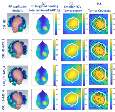 |
131 |
Electric Dipole Based Metasurfaces Enhance Targeted RF Power
Deposition of Thermal Magnetic Resonance at 7.0T, 9.4T and 10.5T
Nandita Saha1,2,
Rita Schmidt3,
and Thoralf Niendorf1,4
1Berlin Ultrahigh Field Facility (B.U.F.F.), Max-Delbrück Center for Molecular Medicine in the Helmholtz Association, Berlin, Germany, Berlin, Germany, 2The Charité – Universitätsmedizin, Berlin, Germany, 3Department of Neurobiology, Weizmann Institute of Science, Rehovot, Israel, 4Experimental and Clinical Research Center (ECRC), a joint cooperation between the Charité Medical Faculty and the Max-Delbrück Center for Molecular Medicine in the Helmholtz Association, Berlin, Germany Keywords: Interventional Devices, RF Arrays & Systems, Metamaterial surface This work proposes the feasibility of passive metasurface (MS) to enhance the RF power deposition for Thermal Magnetic Resonance based treatment of brain tumors. An 8 channel hybrid RF applicator combining dipole antenna and passive MS is designed and evaluated at 7.0 T (300 MHz), 9.4 T (400 MHz) and 10.5 T (450 MHz). MS with metal strips inside waterbolus was designed which generates expected resonant modes of the thermal intervention frequency. The result demonstrated use of passive metamaterial surface in RF applicator enhances the RF power deposition inside target volume for targeted RF heating of deep seated brain tumor. |
|
4393.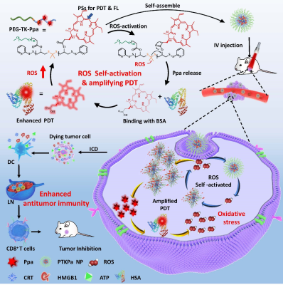 |
132 |
Self-activatable photodynamic-immunotherapy based on
poly(thioketal) conjugated with photosensitizers
Wang Nianhua 1,
Yao wang1,
and Jiang Xinqing1
1Guangzhou First People’s Hospital, Guangzhou, China Keywords: Multimodal, Molecular Imaging, MRI Near Infrared Ray Photodynamic therapy is a non-invasive and controllable modality with potential as a novel cancer treatment strategy. However, the ROS production efficiency of PDT is restricted to hydrophobic characteristics and aggregation caused quenching of PSs. Herein, we designed a ROS self-activatable polymer (PTKPa) to suppress photosensitizers ACQ and elevate ROS production capacity. This platform shows therapeutic outcomes in both cells and orthotopic mouse model, which can significantly elevate the intracellular ROS level upon 660nm laser irradiation, induce immunogenic cell death (ICD) to activate antitumor immunity. This work provides a general strategy enhancing tumor photodynamic therapeutic effects. |
|
4394.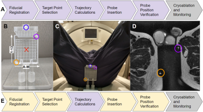 |
133 |
Implementation of Proposed Methods for MR-Guided Transperineal
Prostate Interventions: Real Time Trajectory Calculation and
Monitoring
Thomas Lilieholm1,
David A Woodrum2,
Walter F Block1,3,4,
and Erica M Knavel Koepsel3
1Medical Physics, University of Wisconsin - Madison, Madison, WI, United States, 2Radiology, Mayo Clinic, Rochester, MN, United States, 3Radiology, University of Wisconsin - Madison, Madison, WI, United States, 4Biomedical Engineering, University of Wisconsin - Madison, Madison, WI, United States Keywords: Interventional Devices, Visualization Utilizing MR guidance for minimally-invasive prostate interventions is known to improve the precision and accuracy of the procedure. Previous work has been done to develop and propose an integrated platform for conveniently providing this image guidance to clinicians for use with transperineal prostate interventions. Since then, the proposed methodology has been applied, in a limited capacity, to assist in an in-vivo prostate biopsy. It was found that the trajectory computation component of the platform was able to accurately guide needle placement to the clinician-selected anatomical regions with little need for correction. |
|
4395.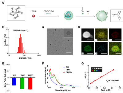 |
134 |
Manganese-Phenolic Networks Platform Amplifying STING Pathway
Activation for MRI Imaging Guided Cancer Chemo- /Immune Therapy
Xinrui Pang1,
Ruili Wei1,
Ye Wang1,
Fangrong Liang1,
and Ruimeng Yang1
1Department of Radiology, Guangzhou First People’s Hospital, GuangZhou, China Keywords: Whole Body, Cancer, Immunity therapy Low immune infiltrates severely hinder cancer immunotherapy. Hence, we developed a manganese-phenolic networks platform (TMPD) The TMPD was based on doxorubicin (DOX) loaded PEG-PLGA nanoparticles and further coated with manganese (Mn)-tannic acid (TA) networks. Mechanistically, DOX could kill cancer cells while Mn2+ could significantly increase the activity of STING pathway related protein to amplify the STING signal and has the function of MRI T1 imaging. The nanoplatform could promote the presentation of tumor antigens and activate a robust innate and adaptive immunity, providing reference for the application of molecular imaging in tumor diagnosis and treatment. |
|
4396.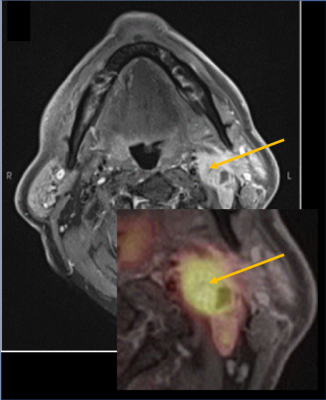 |
135 |
PET-MRI lymph node analysis, a predictive tool for recurrence in
upper aerodigestive tract cancers
Nadya Pyatigorskaya1,
Yoann Sclover2,
Aurelie Kas2,
Lydia Yahia-Cherif1,
and Melika Sahli Amor 2
1ICM, Paris, France, 2APHP, Pitié Salpêtrière, Paris, France Keywords: PET/MR, Head & Neck/ENT, ENT, head and neck, PET/MRI, lymph nodes The aim of our study was to analyze the imaging characteristics of lymph nodes on pre-operative PET-MRI to find the predictors of recurrence or progression.273 patients with ATD cancer who underwent PET-MRI were included. Lymph node PET/MR analysis has shown that the strongest predictors of the recurrence were lymph node enhancement and SUVmax for the controlateral nodes and enhancement, SUVmax node shape for the homolateral nodes. In conclusion, PET-MRI can be an interesting tool in the pre-therapeutic assessment of primary tumors of the ADT with lymph node extension, particularly in the identification of predictive characteristics of recurrence or progression. |
|
4397.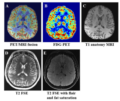 |
136 |
MRI compatibility study of a brain PET insert integrated with a
quadrature birdcage transmit /47-channel receiver head coil
assembly at 3 T
Qiaoyan Chen1,2,
Zhonghua Kuang1,2,
Yongfeng Yang1,2,
Changjun Tie3,4,5,
Xiaoliang Zhang6,
Xin Liu1,2,
Hairong Zheng1,2,
and Ye Li1,2
1Paul C. Lauterbur Imaging Research Center, Shenzhen Institutes of Advanced Technology, Chinese Academy of Sciences, Shenzhen, China, 2Key Laboratory for Magnetic Resonance and Multimodality Imaging of Guangdong Province, Shenzhen, China, 3Institute of Computing Technology, Chinese Academy of Sciences, Beijing, China, 4University of Chinese Academy of Sciences, Beijing, China, 5Peng Cheng Laboratory, Shenzhen, China, 6Department of Biomedical Engineering, State University of New York at Buffalo, New York, NY, United States Keywords: PET/MR, PET/MR Simultaneous positron emission tomography/magnetic resonance imaging (PET/MRI) shows advantages in clinical and research applications for human brain. In this work, MRI compatibility of a brain PET insert integrated with a quadrature birdcage transmit /47-channel receiver head coil assembly was studied. The results of RF field (B1+) distribution, signal-to-noise ratio (SNR) and static field inhomogeneity (ΔB0) show MRI compatibility of the brain PET insert. Simultaneous PET/MRI was implemented, which demonstrated a high performance of the brain PET/MRI system. |
|
4398. |
137 |
3D Printed Alignment Apparatus for Retrofitted Rodent PET-MRI at
9.4 Tesla
Leo Marecki1,
Suyog Pol1,
Robert Zivadinov1,2,
and Ferdinand Schweser1,2
1Buffalo Neuroimaging Analysis Center, Department of Neurology at the Jacobs School of Medicine and Biomedical Sciences, University at Buffalo, The State University of New York, Buffalo, NY, United States, 2Center for Biomedical Imaging, Clinical and Translational Science Institute, University at Buffalo, The State University of New York, Buffalo, NY, United States Keywords: PET/MR, Preclinical We developed a retrofitted alignment apparatus that attaches to the MRI’s motor-driven automated positioning system and enables simultaneous preclinical PET/MRI at 9.4T with a stand-alone microPET ring. The system leverages 3D printing technology to achieve stable alignment between the two modalities, consistent placement of animals, and rapid reconfiguration between PET-MRI and MRI-only experiments. Thereby addressing the shortcomings of other retrofitted designs and proving simultaneous PET-MRI capabilities at a reduced cost. |
|
4399.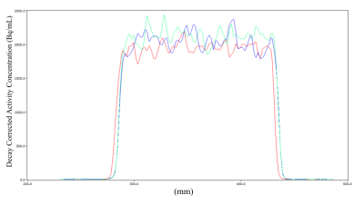 |
138 |
Evaluation of MRI performance and PET compatibility of a 32
channel coil for the Siemens Biograph mMR PET/MRI scanner
Harris Bidaman1,
Katie Dinelle1,
Reggie Taylor2,3,
and Lauri Tuominen2,4
1Brain Imaging Centre, Royal Ottawa Mental Health Centre, Ottawa, ON, Canada, 2University of Ottawa Institute of Mental Health Research at The Royal, Ottawa, ON, Canada, 3Department of Physics, Carleton University, Ottawa, ON, Canada, 4Department of Psychiatry, University of Ottawa, Ottawa, ON, Canada Keywords: PET/MR, Brain, Coil We evaluate MRI and PET performance of the novel Siemens 32 channel head coil for the Siemens Biograph mMR. Results are compared to the system standard 12 channel coil. MRI performance (SNR) was improved with the 32 channel coil. PET image uniformity was not impacted. ROI analysis of the PET data indicated a 5% underestimation of activity concentration in the 32 channel coil, however this difference was mitigated by the use of an image derived reference region. Any small differences in PET performance introduced by the 32 channel coil are far outweighed by the superior MRI performance of this coil. |
|
4400.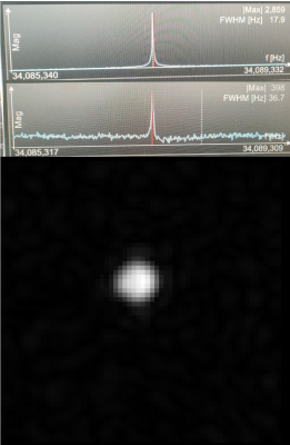 |
139 |
Development of Small Animal PET-insert Compatible RF Coils for
Simultaneous 129Xe MRI/15O2-PET Brain Perfusion Measurements
Elise Noelle Woodward1,
Heeseung Lim2,3,
Ramanpreet Kaur Sembhi1,
Matthew S Fox1,2,
and Alexei Ouriadov1,4
1Physics and Astronomy, Western University, London, ON, Canada, 2Lawson Health Research Institute, London, ON, Canada, 3Siemens Healthcare Limited, London, ON, Canada, 42Lawson Health Research Institute, London, ON, Canada Keywords: RF Arrays & Systems, Hyperpolarized MR (Gas) This study aims to present two birdcage radiofrequency (RF) coils for the use of simultaneous hyperpolarized Xenon-129 (129Xe) MRI and 15O2 PET imaging for brain perfusion measurements. Coils were designed for use with a 2.89T Siemens scanner, or 34.05MHz, one using a low pass filter design, and the other a high pass filter design. |
|
4401.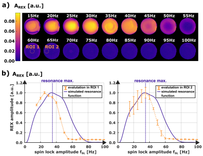 |
140 |
Direct Detection of Peak-like Magnetic Field Fluctuations using
Rotary Excitation (REX) based MRI
Petra Albertova1,2,
Maximilian Gram1,2,
Martin Blaimer3,
Magnus Schindelhütte4,
Thomas Kampf4,
Peter Michael Jakob2,
and Peter Nordbeck1,5
1Department of Internal Medicine I, University Hospital Würzburg, Würzburg, Germany, 2Experimental Physics 5, University of Würzburg, Würzburg, Germany, 3Fraunhofer Institute for Integrated Circuits IIS, Würzburg, Germany, 4Department of Diagnostic and Interventional Neuroradiology, University Hospital Würzburg, Würzburg, Germany, 5Comprehensive Heart Failure Center (CHFC), University Hospital Würzburg, Würzburg, Germany Keywords: Bioeffects & Magnetic Fields, Pulse Sequence Design, spin-lock With spin-locking, the resonance of the spin system can be shifted adjustably to the low frequency range for detection of biomagnetic field oscillations. In this study, we demonstrate that spin-locking is also sensitive to peak-like magnetic field fluctuations with broader spectral width. Simulations and measurements demonstrate that, in addition to the application of imaging neural oscillations, the spatially resolved detection of biomagnetic peaks is possible. Initial results show a high agreement between simulations and experiments and mark a first step towards spin-lock based diagnostics of biomagnetic activity, e.g. imaging of the cardiac conduction system. |
|
The International Society for Magnetic Resonance in Medicine is accredited by the Accreditation Council for Continuing Medical Education to provide continuing medical education for physicians.