Digital Poster
MSK in Motion I: Imaging of Muscle & Tendon
ISMRM & ISMRT Annual Meeting & Exhibition • 04-09 May 2024 • Singapore

Digital Poster
MSK in Motion I: Imaging of Muscle & Tendon
| Computer # | |||
|---|---|---|---|
1530.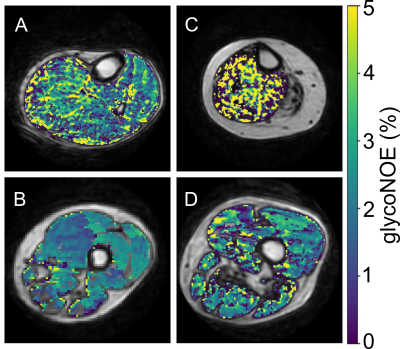 |
129 |
Mapping human skeletal muscle glycogen in Pompe disease patients
Qing Zeng1,2,
Glenn A. Walter3,
Peter C.M. van Zijl1,2,
Manuela Corti4,
Matthew S. Gentry5,6,
Ramon C. Sun5,6,
Barry J. Byrne4,
and Nirbhay N. Yadav1,2
1Russell H. Morgan Department of Radiology and Radiological Science, Johns Hopkins University School of Medicine, Baltimore, MD, United States, 2F.M. Kirby Research Center for Functional Brain Imaging, Kennedy Krieger Institute, Baltimore, MD, United States, 3Department of Physiology and Functional Genomics, University of Florida, Gainesville, FL, United States, 4Department of Pediatrics, University of Florida, Gainesville, FL, United States, 5Department of Biochemistry & Molecular Biology, College of Medicine, University of Florida, Gainesville, FL, United States, 6Center for Advanced Spatial Biomolecule Research, University of Florida, Gainesville, FL, United States Keywords: Muscle, Rare disease, metabolism, molecular imaging Motivation: Pompe disease is a glycogen storage disease which leads to abnormal glycogen accumulation in tissues such as skeletal muscle, but there is a lack of suitable noninvasive methods to assess disease progression and treatment response. Goal(s): To develop an MRI method for assessing glycogen levels in skeletal muscle of Pompe disease patients. Approach: We used glycoNOE MRI to detect glycogen and quantified signals using a Voigt and polynomial hybrid lineshape fitting model. Results: The glycoNOE signals showed significantly higher glycoNOE contrast in skeletal muscle for Pompe patients compared to the control (p < 0.0001). Impact: Glycogen level is an import marker for Pompe disease. Our proposed method is expected to be a useful tool for assessing disease progression and treatment response in Pompe disease and other glycogen storage diseases. |
|
| WITHDRAWN | |||
1532.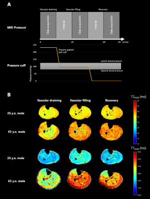 |
130 |
Comparison of water T1 and T2 dynamics in lower extremity
muscles under cuff compression paradigm
Eléonore VERMEULEN1,
Pierre-Yves Baudin1,
Yves Fromes1,
Jean-Marc Boisserie1,
and Benjamin Marty1
1NMR Laboratory, Neuromuscular Investigation Center, Institute of Myology, Paris, France Keywords: Muscle, MSK Motivation: Water-T2 (T2H2O) is a frequently used biomarker of neuromuscular active muscle damage. Complementarity with water-T1 (T1H2O) has been recently suggested, with significant correlations found between T1H2O and T2H2O values in subject with different neuromuscular disorders. Goal(s): In this work, we aimed to explore the behavior of these two biomarkers in the calf muscles of healthy volunteers under different physiological conditions. Approach: T1H2O and T2H2O were measured under different vascular filling conditions expected to modify muscle extracellular volume.
Results: There was a significant correlation between
T1H2O and
T2H2O values
in all conditions. Impact: Water relaxation times T1H2O and T2H2O were measured in the calves of volunteers under different vascular filling conditions. Their distinct behavior suggests that these two biomarkers are complementary in the study of neuromucular diseases. |
|
1533. |
131 |
Calf muscle compartmental T1, T2, T1ρ, and Fat Fraction
associations with Muscle Function in Patients with Heart Failure
Brendan L. Eck1,
Valesha Province2,
Ria Tilve1,
Timothy Engelman2,
W. H. Wilson Tang2,
and Xiaojuan Li1
1Biomedical Engineering, Cleveland Clinic, Cleveland, OH, United States, 2Cardiovascular Medicine, Cleveland Clinic, Cleveland, OH, United States Keywords: Muscle, Muscle, Multiparametric, relaxometry, biomarkers, fat Motivation: Skeletal muscle degeneration (sarcopenia) is more prevalent in patients with heart failure (HF) than the general population and is associated with worsened outcomes. However, HF-associated muscle degeneration remains incompletely understood. Goal(s): To evaluate quantitative MRI in calf muscle compartments of HF patients and to investigate associations with function and overall condition. Approach: Relaxometric (T1/T2/T1ρ) and proton density fat fraction (PDFF) measures were obtained in calf muscle compartments of patients with HF. Functional and survey measures were obtained. Correlation analyses between all measures were performed. Results: Relaxometry and PDFF correlated with muscle function but differed by muscle compartment, suggesting heterogeneous changes as function declines. Impact: Quantitative MRI, including relaxometry and fat fraction as complementary measures, may detect muscle compartmental changes in heart failure associated sarcopenia. Characterization of degradation in patients’ specific muscle compartments may provide insight to the degenerative process and targets for intervention. |
|
1534.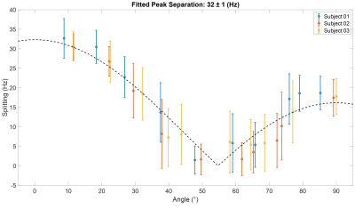 |
132 |
Measuring Quadrupolar Splittings in Human Lower Leg and Forearm
Muscle after Deuterium Oxide Loading
Robin Damion1,2,3,
Daniel Cocking3,4,
Matthew Brook2,5,6,
Dorothee Auer1,2,3,
and Richard Bowtell2,3,4
1School of Medicine, University of Nottingham, Nottingham, United Kingdom, 2NIHR Nottingham Biomedical Research Centre, University of Nottingham, Nottingham, United Kingdom, 3Sir Peter Mansfield Imaging Centre, University of Nottingham, Nottingham, United Kingdom, 4School of Physics, University of Nottingham, Nottingham, United Kingdom, 5School of Life Sciences, University of Nottingham, Nottingham, United Kingdom, 6MRC-Versus Arthritis Centre for Musculoskeletal Ageing Research, University of Nottingham, Nottingham, United Kingdom Keywords: Muscle, Deuterium, Quadrupolar effects Motivation: Although it is known that deuterium (2H) quadrupolar splittings arise in muscle, it is not clear to what extent they can be used to characterise tissues. Goal(s): To investigate quadrupolar splittings by measuring angular dependence and double-quantum-filtered signals, and to determine whether splittings were related to specific muscles. Approach: Deuterium oxide loading was used to increase the 2H signals. The angular dependence of the splitting was measured in the forearm and DQF spectra were acquired on forearm and lower leg muscles. Results: Evidence was obtained for quadrupolar splittings which were shown to depend on angle and muscle group, and exhibited DQF spectra. Impact: Deuterium quadrupolar splittings have the potential to characterise muscle fibres in vivo. Understanding the origin of the splittings, and factors affecting their magnitude, could lead to new or complementary methods in musculoskeletal or physiological investigations. |
|
1535.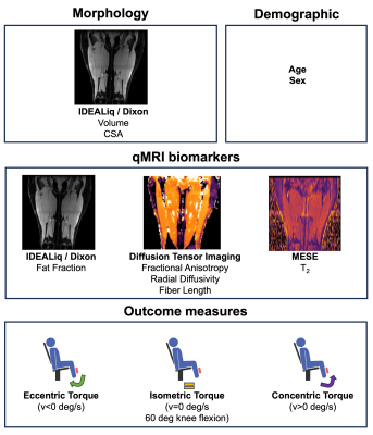 |
133 |
Imaging biomarkers of skeletal muscle strength across the
lifespan
Valentina Mazzoli1,2,
Yael Vainberg2,
Mary E Hall2,
Jessica Asay2,
Scott Delp3,
Feliks Kogan2,
and Garry E Gold2
1Bernard and Irene Schwartz Center for Biomedical Imaging, Department of Radiology, New York University, New York, NY, United States, 2Department of Radiology, Stanford University, Stanford, CA, United States, 3Mechanical Engineering, Stanford University, Stanford, CA, United States Keywords: Muscle, Aging Motivation: Physical therapy and exercise are proposed to slow down the aging-associated loss in muscle strength, but it is currently not known which compositional or architectural aspects of skeletal muscle cause reduced muscle strength. Goal(s): Our goal was to exploit quantitative MRI to identify determinants of muscle strength production across the lifespan. Approach: We used quantitative MRI (fat fraction, diffusion parameters, fiber length and muscle T2) to predict quadriceps torque (n=24, 30-80 y/o). Results: We found that the inclusion of FA greatly improved the prediction of torque over morphology alone. This might be explained with modifications of fiber typing with aging. Impact: We have demonstrated that DTI parameters provide quantitative metrics of muscle quality which can be used to study force production in skeletal muscle, independently of volume. These compositional aspects might be amenable to interventions and provide specific targets for treatment. |
|
1536. |
134 |
Large-scale analysis of subregional thigh muscle volumes on
Dixon MRI and potential clinical utility
Fan Huang1,
Jie Lian1,
and Varut Vardhanabhuti1
1Department of Diagnostic Radiology, The University of Hong Kong, Hong Kong, Hong Kong Keywords: Muscle, Muscle Motivation: Asymmetrical muscle atrophy and hypertrophy are known to occur in muscular atrophy. Musclar subtle changes are important indicators for early detection and tracking changes longitudinally with intervention. Goal(s): To automatically segment thigh muscle subregions and assess their muscular volume and trends with ageing on large-scale data. Approach: We trained nnU-Net for thigh muscle segmentation, and applied on the UK-Biobank data to assess the association between muscular volume and aging and diseases. Results: Thigh musclar volume decreases as we are aging, and is significantly inversely associated with type-2 diabetes, hyptertebnsion and dementia. Impact: Lower volume of the anterior thigh muscle was found in type-2 diabetes, hypertension and dementia patients. Lower volumes of the medial and posterior thigh muscle were found in dementia patients. |
|
1537.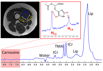 |
135 |
Reproducibility of in vivo measurements of carnosine in muscles
with 1H-MR spectroscopy
Maik Rothe1,2,
Walter Alexander Wohlgemuth1,2,
and Alexander Gussew1,2
1Medical Physics Group, University Clinic and Outpatient Clinic for Radiology, University Hospital Halle (Saale), Halle (Saale), Germany, 2Halle MR Imaging Core Facility, Medical Faculty, Martin-Luther-University Halle-Wittenberg, Halle (Saale), Germany Keywords: Muscle, Spectroscopy Motivation: Due to its pH-buffering function in skeletal muscle, carnosine reveals muscle fibre specific concentrations and is therefore suitable as a marker for the evaluation of muscle fibre composition in pathologies. Goal(s): This study examines the reproducibility of muscle carnosine quantitation with 1H-MRS, and is further targeting to adjust scan parameters for future clinical studies. Approach: 1H-MRS was applied in healthy volunteers to evaluate carnosine lateralization in leg muscles as well as quantitation reproducibility and precision in various parameter settings. Results: Muscular carnosine can be measured with a sufficient precision and reproducibility in less than four minutes and in small voxels. Impact: 1H-MRS enables reliable in vivo measurements of carnosine in skeletal muscles in clinical protocol settings, which is important for the assessment of disease- and age-related as well as myodegenerative changes in muscle fibre composition. |
|
1538.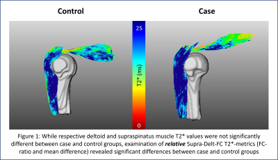 |
136 |
Quantitative MRI Evaluation of the Supraspinatus-Deltoid Muscle
Force Couple with T2* Mapping
Erin C Argentieri1,
Peder E.Z Larson1,
Drew A. Lansdown2,
Brian T. Feeley2,
Sharmila Majumdar1,
and Misung Han1
1Radiology and Biomedical Imaging, University of California San Francisco, San Francisco, CA, United States, 2Department of Orthopaedic Surgery, University of California San Francisco, San Francisco, CA, United States Keywords: Muscle, MSK, UTE, T2* mapping, shoulder, muscle, supraspinatus, deltoid Motivation: No previous studies have evaluated relative supraspinatus-deltoid force couple (Supra-Delt-FC) muscle T2*-metrics or considered their potential as biomarkers for Supra-Delt-FC function Goal(s): Evaluate/compare Supra-Delt-FC T2*-values within 6 supraspinatus injured-case, and 6 (age/sex matched) uninjured-control subjects Approach: Unilateral, 3T, 3D-Cones, 15-echo-UTE MRI acquisitions were utilized for calculation and evaluation of Supra-Delt-FC muscle T2*-values Results: Independent evaluations of deltoid and supraspinatus muscle T2*-values revealed no significant differences between injured-case and uninjured-control subjects. However, evaluation of relative T2*-metrics (mean difference & FC-ratios) revealed significant case-to-control differences – suggesting that relative supraspinatus-to-deltoid T2* metrics may be more sensitive to alterations in Supra-Delt-FC and shoulder function following injury Impact: While independent comparisons of respective supraspinatus and deltoid muscle T2*-values revealed no significant case-to-control differences, relative Supra-Delt-FC muscle T2*-metrics were significantly different between groups. Results suggest that relative Supra-Delt-FC muscle T2*-metrics may be more sensitive to alterations in Supra-Delt-FC function. |
|
1539.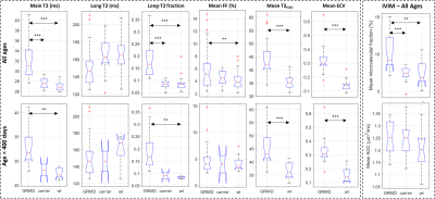 |
137 |
Quantifying tissue water compartmentation for assessing specific
histopathological changes in dystrophic skeletal muscle
Ericky Caldas De Almeida Araujo1,
Inès Barthélémy2,
Yves Fromes1,
Pierre-Yves Baudin1,
Stéphane Blot2,
Harmen Reyngoudt1,
and Benjamin Marty1
1Neuromuscular Investigation Center, Institute of Myology, Paris, France, 2Université Paris Est Créteil, INSERM, IMRB, EnvA, Créteil, France Keywords: Muscle, Relaxometry, Multi-component water T2 Motivation: Developing an objective measure of specific histopathological changes in the skeletal muscle in Duchene Muscular Dystrophy. Goal(s): To investigate distinct histopathological changes in dystrophic muscle by assessing histological tissue water compartmentation in vivo. Approach: A cohort of Golden Retriever Muscular Dystrophy (GRMD), carrier, and wild type dogs underwent quantitative MRI. Tissue water compartmentation, microvascular fraction (f) and extracellular volume (ECV) were assessed using multi-component water-T2 relaxometry, Intravoxel Incoherent Motion (IVIM) and T1 maps acquired before and after gadolinium (Gd) injection. Results: In GRMD, ECV, f, long-T2 fraction and main-T2 value were increased. Carrier dogs displayed mild non-significant abnormalities. Impact: Evaluating tissue water compartmentation in GRMD provided histopathologic specificity, distinguishing vascular from parenchymal changes in dystrophic muscle. The ability to assess histopathologic changes non-invasively will drastically improve our understanding of the disease evolution, treatment strategies and, ultimately, therapeutic development. |
|
1540.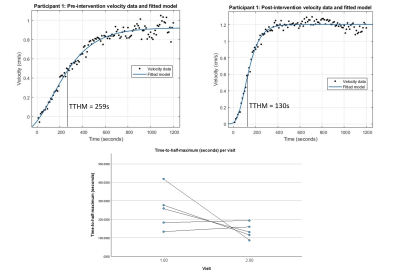 |
138 |
Stimulated phase contrast motor unit MRI to assess recovery of
fatigued muscle as a novel therapeutic response biomarker
Mathew Elameer1,2,
Matthew Birkbeck1,3,
Linda Heskamp1,
Jane Newman4,
Renae Stefanetti4,
Isabel Barrow4,
Gráinne Gorman4,
Ian Schofield1,
Julie Hall2,
Andrew Blamire1,
and Roger Whittaker1
1Translational and clinical research institute, Newcastle University, Newcastle-upon-Tyne, United Kingdom, 2Department of Neuroradiology, Royal Victoria Infirmary, Newcastle-upon-Tyne, United Kingdom, 3Northern Medical Physics and Clinical Engineering, Newcastle upon Tyne Hospitals NHS Foundation Trust, Newcastle-upon-Tyne, United Kingdom, 4Wellcome Centre for Mitochondrial Research, Newcastle University, Newcastle-upon-Tyne, United Kingdom Keywords: Muscle, Neuroscience Motivation: Current biomarkers for serial functional muscle assessments (eg., for therapeutic response assessment) are limited by sensitivity, spatial resolution, and coverage. Goal(s): We aimed to utilise the high spatial resolution offered by recently developed phase contrast motor unit MRI (PC-MUMRI) techniques to identify potential biomarkers. Approach: We prospectively trialled a novel PC-MUMRI fatigue and recovery paradigm before and after a 12-week exercise intervention in seven participants with genetic mitochondrial disorders. Results: Time-to-half-maximum of PC-MUMRI recovery reduced from a mean of 254 (+/- 109) seconds to 137 (+/- 41) seconds following the intervention. This was not statistically significant (p = 0.074). Impact: We have developed and tested a novel therapeutic response biomarker for muscle-based intervention based on measuring recovery of stimulated muscle twitch velocities following fatigue. This may address problems with spatial resolution, sensitivity, or coverage associated with previously reported biomarkers. |
|
1541. |
139 |
Texture analysis of T1 and T2 weighted images allows to identify
STZ-induced diabetic sarcopenia rats with skeletal muscle fiber
atrophy
Dong Xing1 and
Yunfei Zha1
1Department of Radiology, Renmin Hospital of Wuhan University, Wuhan, China Keywords: Muscle, Muscle Motivation: Looking for a convenient tool to detect the skeletal muscle fiber atrophy non-invasively in the early stage of diabetic sarcopenia. Goal(s): This study aims to assess the pathological changes in skeletal muscle of diabetic sarcopenia rats using muscle MR texture analysis. Approach: Type 1 diabetic sarcopenia rat model was conducted via STZ intraperitoneal injection. 2D MR texture features of the gastrocnemius muscle were extracted, followed by histopathological examination using HE stains. Results: Muscle MR texture features exhibited significant differences between the diabetic sarcopenia group and the control group, and demonstrated a strong correlation with pathological characteristics of skeletal muscle fibers. Impact: Texture features derived from muscle MR images show potential for non-invasively identifying skeletal muscle atrophy in diabetic sarcopenic rats and providing insights into underlying disease mechanisms. |
|
1542.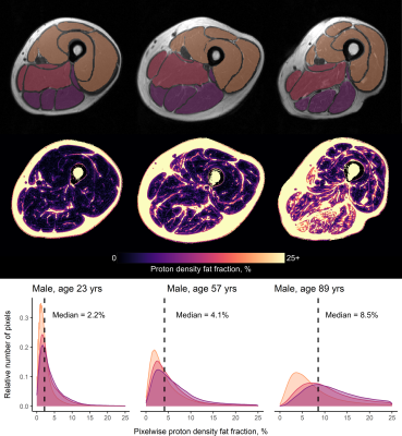 |
140 |
Skeletal muscle involvement in ageing: Exploring the
relationship between MRI fat fraction and age in the muscles of
the thigh
Jamie Scott1,
David A. Reiter2,
Fatemeh Adelnia3,
Christopher M. Bergeron4,
Kenneth W. Fishbein4,
Max Yates1,
Richard G. Spencer4,
Ailsa A. Welch1,
Luigi Ferrucci5,
and Donnie Cameron1,5,6
1Norwich Medical School, University of East Anglia, Norwich, United Kingdom, 2Emory University School of Medicine, Atlanta, GA, United States, 3Vanderbilt University Institute of Imaging Science, Nashville, TN, United States, 4Laboratory of Clinical Investigation, National Institute on Aging, Baltimore, MD, United States, 5Translational Gerontology Branch, National Institute on Aging, Baltimore, MD, United States, 6Department of Medical Imaging, Radboud University Medical Center, Nijmegen, Netherlands Keywords: Muscle, Aging, Sarcopenia, muscle quality, fat replacement
Motivation: Deposition of fat in skeletal muscle
increases with age, leading to reduced muscle quality, but
it is currently unclear which muscles are affected first and
thus may serve as markers for the onset of this process. Goal(s): To measure intramuscular fat in individual thigh muscles in a healthy ageing cohort. Approach: We applied chemical-shift-based water-fat-separation imaging in 94 participants (median age=56, range=22-89yrs), and proton density fat fraction was calculated for 12 thigh muscles and different muscle groups.
Results: We showed age associations with fat
deposition in the whole thigh overall (β=0.60, p≪0.001),
with associations being stronger in women and in the
hamstring muscles. Impact: Understanding the relationship between proton density fat fraction and age in the thigh musculature—particularly in women and in the hamstring muscles—will help clinicians to identify specific muscle targets for interventions designed to reduce functional decline with ageing. |
|
1543.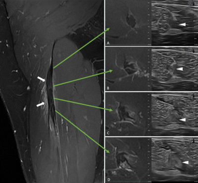 |
141 |
Improving the assessment of myotendinous injuries on
professional athletes using Ultrasound-MRI fusion exploration
Manuel Wong1,
Sandra Mechó1,2,
Priscilla Jarrin1,
Alicia Palomar García3,
Valentin H. Prevost4,
Wolter L. de Graaf5,
and Xavier Yanguas1
1Medical Department of Futbol Club Barcelona (FIFA Medical Center of Excellence), Barcelona, Spain, 2Department of Radiology, Hospital de Barcelona, SCIAS, Barcelona, Spain, 3Canon Medical Systems Spain and Portugal, Barcelona, Spain, 4Canon Medical Systems Corporation, Tochigi, Japan, 5Canon Medical Systems Europe, Amstelveen, Netherlands Keywords: Tendon/Ligament, Multimodal, Ultrasound-MRI fusion, myotendinous injury, sports medicine, injury prognosis, lesion follow-up Motivation: US-MRI fusion could provide more accurate information for muscle injury evaluation and follow-up. To date, some works were conducted in the scope of prostate pathology, but only a few focused on musculoskeletal imaging. Goal(s): This study aimed to evaluate if US-MRI fusion would allow more detailed and precise characterization of myotendinous injuries. Approach: US only and US-MRI fusion images were reviewed and scored in terms of muscle injury detection, as well as scar, tendon and edema visualization. Results: US-MRI fusion allowed a better characterization of lesions and healing process on professional athletes suffering from myotendinous injuries compared to US alone. Impact: Diagnosis of muscle injuries by US is a challenge, so MRI is still the gold standard in most cases. Combining both techniques, we can better characterize and follow-up lesions, detecting a slowdown in healing or complications as soon as possible. |
|
1544.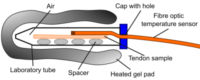 |
142 |
Temperature dependence of T1 and T2* in ex vivo ovine Achilles
tendons
Marta B. Maggioni1,
Matthias M. Kollert2,
Nicholas M. Brisson2,
Georg N. Duda2,
Jürgen R. Reichenbach1,
and Martin Krämer1,3
1Medical Physics Group, Institute of Diagnostic and Interventional Radiology, Jena University Hospital, Friedrich Schiller University, Jena, Germany, 2Julius Wolff Institute, Berlin Institute of Health at Charité, Universitätsmedizin, Berlin, Germany, 3Institute of Diagnostic and Interventional Radiology, Jena University Hospital, Friedrich Schiller University, Jena, Germany Keywords: Tendon/Ligament, Quantitative Imaging, Temperature Motivation: While the dependence of the relaxation parameters T1 and T2* on temperature has been studied in aqueous solutions and in some biological tissues, such observations for complex organized tissues including tendons have not been investigated previously. Goal(s): To measure how T1 and T2* change with temperature in Achilles tendon samples. Approach: T1 and T2* were continuously measured while fresh ex vivo Achilles tendon samples were placed in a heated environment and cooled down over several hours. Results: The change in T2* with temperature was nonlinear and much higher than that reported in the literature for other tissues Impact: The change in T2* in Achilles tendons was 4.8+/-2.1%/°C, which is substantially higher than reported in other biological tissues and indicates that the theory of temperature dependence of MRI relaxation parameters may not apply to highly organized tissues like tendons. |
|
1545.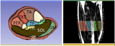 |
143 |
Progression of Miyoshi muscular dystrophy monitored by
quantitative MRI
Ivica Just1,2,
Petra Hnilicova3,
Radka Klepochova2,4,
Siegfried Trattnig5,6,7,8,
Monika Turcanova Koprusakova9,
Martin Kolisek3,
and Martin Krššák4
1Internal Medicine III, Medical University of Vienna, Vienna, Austria, 2High Field MR Center, Department of Biomedical Imaging and Image-Guided Therapy, Medical University of Vienna, Vienna, Austria, 3Biomedical Centre Martin, Comenius University in Bratislava, Jessenius Faculty of Medicine in Martin, Martin, Slovakia, 4internal Medicine III, Medical University of Vienna, Vienna, Austria, 5High Field MR Center, Department of Biomedical Imaging and Image-Guided Therapy, Medical University of Vienna, Vienna, Austria, 6CD Laboratory for MR Imaging Biomarkers (BIOMAK), Vienna, Austria, 7Austrian Cluster for Tissue Regeneration, Ludwig Boltymann Institute for Experimental and Clinical Traumatology, Vienna, Austria, 8Institute for Clinical Molecular MRI in the Musculoskeletal System, Karl Landsteiner Society, Vienna, Austria, 9Department of Neurology, Comenius University in Bratislava, Jessenius Faculty of Medicine in Martin, Martin, Slovakia Keywords: Muscle, Rare disease, dystrophy, dysferlinopathy Motivation: To characterize the skeletal muscle and assess progression of the Miyoshi dystrophy by quantitative MRI. Goal(s): Compare the changes in fat fractions in individual calf muscles 10 months apart in time. Approach: Lower extremity of 4 dystrophic patients were measured in 3T scanner by multiecho-Dixon sequence in two time points. Three asymptomatic DYSF gene carriers served as reference. Results: Fat fraction increased in all measured muscles in dystrophic patients while staying stable in controls. The most significant increments were detected in the muscles most preserved (TP, FDL, EXT, FHL). Variation can be observed even within the patients with the same genotype. Impact: Using quantitative analysis based on automatically generated fat fraction maps for assessment of Miyoshi dystrophy in muscles allow clinically feasible monitoring of the disease progression and description of the pattern of the disease. |
|
 Back to Meeting Home
 Back to the Program-at-a-Glance
The International Society for Magnetic Resonance in Medicine is accredited by the Accreditation Council for Continuing Medical Education to provide continuing medical education for physicians.
 View Presentation Video