Weekday Course
Forensic & Histology MRI: Bridging Physics, Biology & Pathology
ISMRM & ISMRT Annual Meeting & Exhibition • 04-09 May 2024 • Singapore

Weekday Course
Forensic & Histology MRI: Bridging Physics, Biology & Pathology
| 13:45 |
Forensic Brain MRI
Claudia Lenz, Eva Scheurer
Keywords: Neuro: Brain Forensic medicine employs scientific disciplines to solve legal queries, with forensic imaging, including MRI, as its newest branch. Postmortem cases are investigated to clarify the cause and manner of death, to reconstruct violent events, to determine the time of death and the presence of diseases. Postmortem MRI faces challenges such as temperature variations, postmortem changes and formalin fixation effects. This talk gives an overview on the advantages and challenges of in situ and ex situ postmortem examinations. It emphasizes methods for adapting MRI techniques for forensic brain MRI, highlighting the current research for forensic purposes and the validation of biomarkers. |
|
| 14:15 |
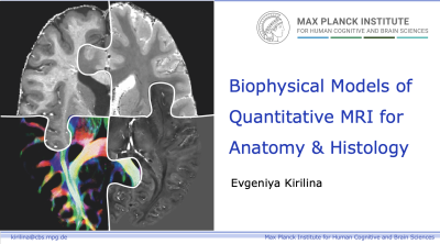 |
Biophysical Models of Quantitative MRI for Anatomy & Histology
Evgeniya Kirilina
Keywords: Neuro: Brain, Contrast mechanisms: Relaxometry Quantitative magnetic resonance imaging (qMRI) expands upon traditional MRI by providing specific physical parameters, like proton density, relaxation rates, diffusion properties of water nuclear spins. These parameters reveal details about local microstructural environments (e.g., brain myelin and iron). Non-invasive in vivo histology via MRI (hMRI) seeks to directly characterize tissue microstructure, potentially replacing or complementing invasive histology. Understanding tissue contrast from hMRI requires biophysical models. This talk explores concepts, models, and validation methods in this area, highlighting challenges and recent progress with the focus on myelin and iron-induced contrasts in the human brain. |
| 14:45 |
Forensic MRI of the Whole Body: Challenges & Opportunities
Eva Scheurer, Claudia Lenz
Keywords: Neuro: Brain TBA |
|
| 15:15 |
UTE for Fracture Detection & Age Evaluation
Jiang Du
Keywords: Musculoskeletal: Skeletal, Image acquisition: Quantification, Image acquisition: Sequences This lecture talks about recent technical developments in ultrashort echo time (UTE) magnetic resonance imaging and applications in fracture detection and age evaluation. A series of techniques have been developed for high contrast imaging of cortical and trabecular bone. Quantitative UTE techniques have also been developed for mapping of T1, T2*, magnetization transfer ratio (MTR), MT modeling of macromolecular fraction (MMF), quantitative susceptibility mapping (QSM) of bone susceptibility, as well as total, bound, and free water in bone. Applications in fraction detection and age evaluation are also discussed. |
 Back to Meeting Home
 Back to the Program-at-a-Glance
The International Society for Magnetic Resonance in Medicine is accredited by the Accreditation Council for Continuing Medical Education to provide continuing medical education for physicians.
 View Presentation Video