Oral
Neuroinflammation: Follow-Up from 2023 Clinical Focus Meeting
ISMRM & ISMRT Annual Meeting & Exhibition • 04-09 May 2024 • Singapore

Oral
Neuroinflammation: Follow-Up from 2023 Clinical Focus Meeting
| 15:45 |
1046.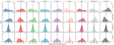 |
Distinct virtual histology of gray matter atrophy in
neuroinflammatory diseases
Jun Sun1,
Yuerong Lizhu1,
Min Guo1,
Siyao Xu1,
Xianchang Zhang2,
Zhizheng Zhuo1,
and Yaou Liu1
1Capital Medical Universtiy, Beijing Tiantan Hospital, Beijing, China, 2MR Research Collaboration, Siemens Healthineers Ltd., Beijing, China Keywords: Neuroinflammation, Neuroinflammation, virtual histology Motivation: Gray matter (GM) atrophied early in multiple sclerosis (MS), anti-aquaporin-4 antibody-positive [AQP4+] / -negative [AQP4-] neuromyelitis optica spectrum disorder (NMOSD), and myelin oligodendrocyte glycoprotein antibody-associated disease (MOGAD). Their neurobiological underpinnings have not been clarified. Goal(s): The purpose was to explore their GM atrophy-associated histology using a multicenter cohort. Approach: 324 MS, 197 AQP4+ NMOSD, 75 AQP4- NMOSD, 47 MOGAD, and 2,169 healthy controls (HCs) were examined by virtual histology method. Results: The unique virtual histology was glial cells for MS, astrocytes for AQP4+ NMOSD and oligodendrocytes for MOGAD. The neuronal and endothelial cells were shared potential targets. Impact: It might help to optimize therapy. |
| 15:57 |
1047.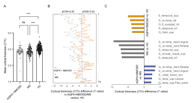 |
Identification of transcriptomic signatures associated with
cortical thinning in neuromyelitis optica spectrum disorder
Min Guo1,
Zhizheng Zhuo1,
Minghao Wu1,
Jun Sun1,
Yu-Xin Yang2,
Yunyun Duan1,
and Yaou Liu1
1Department of Radiology, Beijing Tiantan Hospital, Capital Medical University, Beijing, China, 2United Imaging Research Institute of Intelligent Imaging, Beijing, China Keywords: Neuroinflammation, Genetics Motivation: Determining the potential transcriptomic signatures driving cortical thinning in neuromyelitis optica spectrum disorder (NMOSD) and multiple sclerosis (MS). Goal(s): To compare the cortical atrophy patterns and the underlying molecular mechanisms between NMOSD and MS. Approach: A partial least squares (PLS) regression model was used to associate the CTh alteration profile with the expression of genes from the Allen Human Brain Atlas (AHBA) database, then Metascape analysis was performed to identify the functional biological processes. Results: Distinct cortical atrophy patterns and underlying cell signaling pathways were observed in NMOSD and MS. Impact: The distinct cortical atrophy patterns will help in differential diagnosis of NOMOSD and MS. The identified genes and signaling pathways will help understand the pathological mechanism of both diseases and provide potential therapeutic targets. |
16:09 |
1048.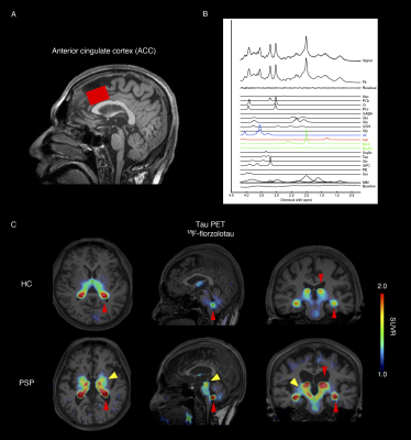 |
In vivo assessment of astrocyte reactivity in patients with
progressive supranuclear palsy
Kosei Hirata1,2,
Kiwamu Matsuoka1,
Kenji Tagai1,
Hironobu Endo1,
Harutsugu Tatebe1,
Maiko Ono1,
Naomi Kokubo1,
Yuko Kataoka1,
Asaka Oyama1,
Hitoshi Shinoto1,
Keisuke Takahata1,
Takayuki Obata1,
Masoumeh Dehghani3,
Jamie Near3,4,
Kazunari Kawamura1,
Ming-Rong Zhang1,
Hitoshi Shimada1,5,
Hiroshi Shimizu5,
Hiroshi Shimizu5,
Takanori Yokota2,
Takahiko Tokuda1,
Makoto Higuchi1,
and Yuhei Takado1
1National Institutes for Quantum Science and Technology, Chiba, Japan, 2Tokyo Medical and Dental University, Tokyo, Japan, 3Sunnybrook Research Institute, Tronto, ON, Canada, 4University of Toronto, Tronto, ON, Canada, 5Niigata University, Niigata, Japan Keywords: Dementia, Neuro, magnetic resonance spectroscopy, progressive supranuclear palsy, astrocyte reactivity Motivation: Although astrocytic pathology is a pathological hallmark of progressive supranuclear palsy (PSP), the role of astrocytes in the pathophysiology of PSP is not fully understood. Goal(s): This study aimed to evaluate astrocyte reactivity in vivo in patients with PSP. Approach: Astrocyte reactivity was assessed by magnetic resonance spectroscopy and plasma biomarkers, which were verified via tau-PET and histopathological analysis. Results: Our results suggest that, in the anterior cingulate cortex, astrocyte reactivity precedes pronounced tau deposition and neurodegenerative processes and modulates brain function in PSP. Elevated myo-inositol was associated with high lactate levels, suggesting a link between reactive astrocytes and brain energy metabolism changes. Impact: This study assessed astrocyte reactivity in vivo using magnetic resonance spectroscopy and plasma biomarkers, providing insights into the involvement of astrocytes in the pathogenesis of progressive supranuclear palsy. |
| 16:21 |
1049.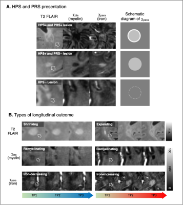 |
Association of Iron Deposition in Early-Stage Multiple Sclerosis
Lesion with Remyelination Capacity: chi-separation imaging study
Hyeong-Geol Shin1,2,
Woojun Kim3,
Hyun-soo Lee4,
Jiwoong Kim5,
Yoonho Nam6,
Xu Li1,2,
Peter van Zijl1,2,
Jongho Lee7,
and Jinhee Jang8
1Radiology, Johns Hopkins University, Baltimore, MD, United States, 2Kennedy Krieger Institute, Baltimore, MD, United States, 3Neurology, Seoul St. Mary's Hospital, Seoul, Korea, Republic of, 4Siemens Healthineers, Seoul, Korea, Republic of, 5Mathematics and Statistics, University of South Florida, Tampa, FL, United States, 6Biomedical Engineering, Hankuk University of Foreign Studies, Yongin, Korea, Republic of, 7Electrical and Computer Engineering, Seoul National University, Seoul, Korea, Republic of, 8Radiology, Seoul St. Mary's Hospital, Seoul, Korea, Republic of Keywords: Multiple Sclerosis, Multiple Sclerosis, 𝜒-separation Motivation: In multiple sclerosis (MS) lesion, factors influencing myelin dynamics and future remyelination are under active investigation. Goal(s): To assess dynamic changes of MS lesions from their early stage and explore the factors related to their future remyelination outcomes.
Approach: Longitudinal changes of MRI phenotypes in
MS lesions were assessed from their early stage,
particularly focusing on longitudinal alternations
in diamagnetic myelin and paramagnetic iron signals using
susceptibility source-separation (chi[𝜒]-separation). Results: 26 lesions show remyelination and 36 did not. The hyperintensity in paramagnetic iron signal (hyper-paramagnetic sign, HPS) at early stage of lesion development was significantly associated to future remyelination. Impact: Iron deposition sign in early-stage MS lesion, which detected by added sensitivity of 𝜒-separation to iron and myelin, can offer potential imaging marker for the impaired remyelination capability in MS pathology. |
16:33 |
1050.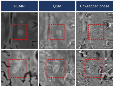 |
Multiparametric Quantitative MRI Shows Enhanced Degeneration in
Paramagnetic Rim Lesions and Their Perilesional Tissue in
Multiple Sclerosis
Alessandro Cagol1,2,3,4,
Mario Ocampo-Pineda1,2,3,
Batuhan Ayci1,5,
Pascal Benkert6,
Po-Jui Lu1,2,3,
Matthias Weigel1,2,3,7,
Lester Melie-Garcia1,2,3,
Xinjie Chen1,2,3,
Antoine Lutti8,
Thanh D. Nguyen9,
Yi Wang9,
Jongho Lee10,
Jens Kuhle2,3,
Ludwig Kappos1,2,3,
Maria Pia Sormani4,
and Cristina Granziera1,2,3
1Translational Imaging in Neurology (ThINk) Basel, Department of Biomedical Engineering, University Hospital Basel and University of Basel, Basel, Switzerland, 2Department of Neurology, University Hospital Basel, Basel, Switzerland, 3Research Center for Clinical Neuroimmunology and Neuroscience Basel (RC2NB), University Hospital Basel and University of Basel, Basel, Switzerland, 4Department of Health Sciences, University of Genova, Genova, Italy, 5Cerrahpasa Medical School, Istanbul University-Cerrahpasa, Istanbul, Turkey, 6Department of Clinical Research, University Hospital Basel, University of Basel, Basel, Switzerland, 7Division of Radiological Physics, Department of Radiology, University Hospital Basel, Basel, Switzerland, 8Laboratory for Research in Neuroimaging, Department of Clinical Neuroscience, Lausanne University Hospital and University of Lausanne, Lausanne, Switzerland, 9Department of Radiology, Weill Cornell Medical College, New York, NY, United States, 10Laboratory for Imaging Science and Technology, Department of Electrical and Computer Engineering, Seoul National University, Seoul, Korea, Republic of Keywords: Multiple Sclerosis, Multiple Sclerosis, Paramagnetic rim lesions; quantitative MRI
Motivation: Paramagnetic rim lesions (PRLs), a subset
of chronic active lesions identifiable through
susceptibility-based imaging, are linked to insidious
disease progression in multiple sclerosis (MS). However,
data on local microstructural changes in PRLs remain
limited.
Goal(s): To comprehensively characterize pathological
alterations within PRLs and the surrounding perilesional
tissue.
Approach: Employing multiparametric quantitative 3T
MRI on 175 people with MS, we obtained contrasts sensitive
to tissue microstructural damage.
Results: PRLs exhibited more pronounced pathological
alterations compared to other white matter lesions,
displaying enhanced demyelination, neuro-axonal loss, and
iron accumulation. Remarkably, these alterations extended
into the perilesional tissue appearing normal on
conventional MRI. Impact: In people with multiple sclerosis, paramagnetic rim lesions (PRLs) exhibit pronounced microstructural quantitative MRI alterations. This strengthens PRLs as reliable biomarkers for lesions with smoldering degenerative activity, and offers potential insights into their association with a more severe disease course. |
| 16:45 |
1051.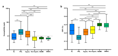 |
The changes of oxygen extraction fraction in different types of
lesions in multiple sclerosis: A cross-sectional and follow-up
study
Yan Xie1 and
Wenzhen Zhu1
1Tongji Hospital, Tongji Medical College, Huazhong University of Science and Technology, Wuhan, China Keywords: Multiple Sclerosis, Multiple Sclerosis Motivation: Multiple sclerosis (MS) lesions with different pathologic conditions could be distinguished by MRI. There may be differences in oxygen metabolism in different types of lesions. Goal(s): To explore the oxygen metabolism of different types of lesions in MS patients by oxygen extraction fraction (OEF) both cross-sectionally and longitudinally. Approach: The OEF map was reconstructed from a 3D multi-echo gradient echo scan. White matter lesions were classified into four types based on contrast-enhanced T1WI and quantitative susceptibility mapping. Results: There were differences in OEF among different types of MS lesions. The OEF in the lesion and the lesion type may change as time progresses. Impact: This study revealed tissue damage and oxygen metabolism level in different types of MS lesions. The OEF may contribute to further understanding of the pathological mechanisms in MS lesion evolution. |
| 16:57 |
1052.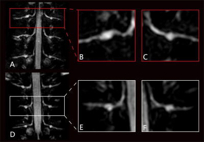 |
Postherpetic Neuralgia and Morphological Alterations in the
Human Dorsal Root Ganglia: A Study Using 3T MR Neurography
Dejun She1,
Yalan Yan1,
Xiance Zhao2,
and Dairong Cao1
1The First Affiliated Hospital of Fujian Medical University, Fuzhou, China, 2Philips Healthcare, Shanghai, China, Shanghai, China Keywords: Neuroinflammation, Neuroinflammation Motivation: Despite the crucial role of imaging in localizing lesions, excluding causes, and guiding surgery, there remains a paucity of reports pertaining to the radiological manifestations in patients with herpetic neuralgia. Goal(s): To investigate the correlation between the volume and T2 signal intensity (SI) of the ganglia in 3T MRN with clinical and serological parameters in herpetic neuralgia patients. Approach: With 18 patients examined by MRN. Volume, T2 signal measurements of the T1-T12 ganglia were performed manually for each patient. Results: The changes in ganglion volume observed on MRN may reflect disease progression. Among all serological indicators, ESR was correlated with the volume ratio. Impact: This study is the first to quantify ganglia in herpetic neuralgia patients, and the first to investigate the correlation between ganglia volume and serological data. In herpetic neuralgia patients, these MRN findings will contribute to the diagnosis and management. |
| 17:09 |
1053.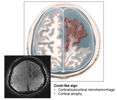 |
Cortical Microhemorrhage Presentation of Small Vessel Primary
Angiitis of the Central Nervous System
Ai Guo1,
Zhe Zhang1,
Ge-Hong Dong1,
Yuan Li2,
Lei Su3,
Chenyang Gao3,
Mengting Zhang1,
Xiaoyu Shi1,
Huabing Wang1,
Xinghu Zhang1,
De-Hong Lu1,
Ying Fu4,
Jing Jing1,
Fu-Dong Shi1,
and De-cai Tian1
1Beijing Tiantan Hospital, Beijing, China, 2MR research collaboration team, siemens healthineers, Beijing, China, 3Tianjin General Hospital, Tianjin, China, 4The First Affiliated Hospital of Fujian Medical University, Fuzhou, China Keywords: Neuroinflammation, Neuroinflammation Motivation: PACNS entails a biopsy for diagnosis, but only with an intermediate sensitivity. It is necessary to revisit PACNS with advanced imaging technique to provide a non-invasive diagnostic standard. Goal(s): We aim to find more pathological details with enough sensitivity and specificity to provide potential biomarkers for PACNS. Approach: 21 patients with small-vessel PACNS were included in this study. T1-MPRAGE, T2 T2*W, and SWI images were collected. Results: Our study highlighted the image features of patients with small-vessel PACNS with coral-like signs through 7T MRI. Due to the small patient cohort, no specific clinical differences between hemorrhagic and non-hemorrhagic patients were found. Impact: The signal characteristics of the coral-like sign represent cerebral cortical microhemorrhages with atrophy, which could be an important MRI pattern of small-vessel PACNS. |
|
17:21 |
1054.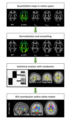 |
COVID-19-related anosmia is driven by inflammation and myelin
alterations as shown by Voxel Based Analysis
Eleonora Lupi1,
Marta Gaviraghi1,
Elena Grosso1,
Anita Monteverdi2,
Marco Battiston3,
Francesco Grussu3,4,
Baris Kanber3,5,
Ferran Prados Carrasco3,5,6,
Janine Makaronidis7,8,
Rebecca S Samson3,
Marios C Yiannakas3,
Egidio D’Angelo1,2,
Fulvia Palesi1,2,
and Claudia A. M. Gandini Wheeler-Kingshott1,2,3
1Department of Brain & Behavioral Sciences, University of Pavia, Pavia, Italy, 2Digital Neuroscience Centre, IRCCS Mondino Foundation, Pavia, Italy, 3NMR Research Unit, Queen Square MS Centre, Department of Neuroinflammation, UCL Queen Square Institute of Neurology, Faculty of Brain Sciences, University College London, London, United Kingdom, 4Radiomics Group, Vall d’Hebron Institute of Oncology, Vall d’Hebron Barcelona Hospital Campus, Barcelona, Spain, 5Department of Medical Physics and Biomedical Engineering, Centre for Medical Image Computing (CMIC), University College London, London, United Kingdom, 6E-Health Center, Universitat Oberta de Catalunya, Barcelona, Spain, 7Centre for Obesity Research, Department of Medicine, University College London, London, United Kingdom, 8National Institute of Health Research, UCLH Biomedical Research Centre, London, United Kingdom Keywords: White Matter, COVID-19, Neuroinflammation, Voxel Based Analysis, g-ratio Motivation: Voxel Based Analysis (VBA) can be a powerful tool to detect localized alterations. Goal(s): Here VBA was used to understand the underpinnings of COVID-19-related anosmia. Approach: Quantitative magnetization transfer and diffusion-weighted imaging derived maps were used to detect pathological changes affecting white matter structures. Results: Microstructural differences were detected between healthy controls and subjects experiencing anosmia or those who recovered from it. Results highlighted the presence of widespread inflammation in persistent anosmia subjects, with myelin damage and possible repair in those who recovered. Myelin alterations involved the olfactory circuit, as well as other brain regions, providing insights into possible mechanisms of COVID-19-related anosmia. Impact: Voxel Based Analysis is a powerful tool to highlight local tissue disruption linked to neuroinflammatory processes. Here VBA provided an insight into microstructure and myelin changes associated to COVID-19-related persistent or recovered anosmia symptoms. |
| 17:33 |
Discussion
Anja van der Kolk
Radboudumc Nijmegen, Netherlands
|
 Back to Meeting Home
 Back to the Program-at-a-Glance
The International Society for Magnetic Resonance in Medicine is accredited by the Accreditation Council for Continuing Medical Education to provide continuing medical education for physicians.
 View Presentation Video