Oral
Psychosis & Mood Disorders
ISMRM & ISMRT Annual Meeting & Exhibition • 04-09 May 2024 • Singapore

| 13:45 |
Introduction
John Port
Mayo Clinic, United States
|
|
| 13:57 |
1239.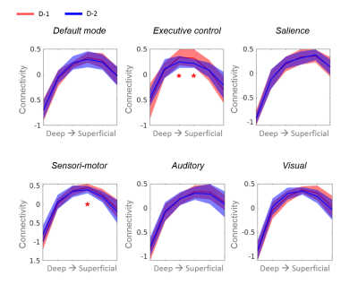 |
Resting-state changes along the cortical depth detected during
evolution of major depressive disorder
Patricia Pais-Roldán1,
Seong Dae Yun1,
Shukti Ramkiran1,2,
Ravichandran Rajkumar1,2,3,
Jana Hagen2,
Areej Al Okla1,
Tanja Veselinovic1,2,
Gereon Schnellbächer2,
Irene Neuner*1,2,3,
and N. Jon Shah*1,3,4,5
1Institute of Neuroscience and Medicine 4, INM-4, Forschungszentrum Jülich, Jülich, Germany, 2Department of Psychiatry, Psychotherapy and Psychosomatics, RWTH, Aachen, Germany, 3JARA - BRAIN - Translational Medicine, Aachen, Germany, 4Department of Neurology, RWTH Aachen University, Aachen, Germany, 5Institute of Neuroscience and Medicine 11, INM-11, JARA, Forschungszentrum Jülich, Jülich, Germany Keywords: Psychiatric Disorders, Brain Connectivity, Laminar connectivity, depression
Motivation: The relative contribution of each
cortical depth to the network disturbances coupled with
depression remains unexplored.
Goal(s): We aim to identify changes in connectivity
along the cortical thickness during depression recovery.
Approach: We use high-resolution, large-coverage EPIK
to functionally map the brain of patients before and after
treatment and perform cortical-depth specific analysis of
network connectivity.
Results: Changes in connectivity linked to depression
treatment were observed at multiple depths of the cortex in
two resting-state networks. Impact: The demonstration that network connectivity does not change homogeneously in a patient population along the cortical depth suggests the importance of adding this variable to the analysis of psychiatric disorders to improve understanding of the mechanisms behind network disturbances. |
14:09 |
1240.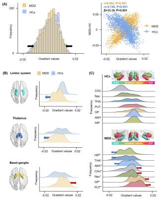 |
Cognition-related connectome gradient dysfunctions of thalamus
and basal ganglia in drug-naïve first-episode major depressive
disorder
Qian Zhang1,
Youjin Zhao1,
Aoxiang Zhang1,
Lizhou Chen1,
and Qiyong Gong1
1Huaxi MR Research Center (HMRRC), Department of Radiology, West China Hospital of Sichuan University, Chengdu, China Keywords: Psychiatric Disorders, Psychiatric Disorders, major depressive disorder, fMRI, functional gradient, subcortical structure, cognition Motivation: The continuous spatial patterns of inter-region connectivity within the subcortical network still remain less well-understood in MDD.
Goal(s): Using functional gradient mapping, a novel
approach to identify hierarchical organization of functional
networks, we aim to evaluate multiscale subcortical
gradients in MDD and their association with cognition. Approach: Subcortical gradient alterations at the global-, system-, and subregion-levels and their relation to neuropsychological functioning were assessed in MDD patients relative to healthy controls. Results: Principal gradient values were lower in thalamic and limbic systems but higher in basal ganglia in MDD. Interactions between thalamic and basal ganglia gradient alterations were implicated in MDD-related memory impairments. Impact: Multiscale subcortical gradient alterations can enhance our understanding of MDD-related hierarchical disturbances in subcortical function and may provide useful clinical biomarkers for cognitive impairments in MDD. |
| 14:21 |
1241. |
Disrupted brain functional connectome gradient in social anxiety
disorder
Xun Zhang1 and
Qiyong Gong1
1Huaxi MR Research Center (HMRRC), West China Hospital of Sichuan University, Chengdu, China Keywords: Functional Connectivity, fMRI (resting state), biomarker Motivation: Although discrete macroscale functional network abnormalities were observed in social anxiety disorder (SAD), the alterations of continuous spatial patterns of functional connectomes remain unknown. Goal(s): We aimed to use a novel method to characterize aberrant patterns of connectome gradients in SAD patients. Approach: We applied diffusion map embedding to characterize functional connectome gradients and investigated between-group differences of global and regional topological features and their clinical relevance. Results: Globally, SAD patients demonstrated decreased explanation ratio, narrower gradient range and less spatial variation in the principal gradient. Regionally, SAD group showed increased gradients in the sensorimotor networks and decreased in the default mode network. Impact: Our findings of internally clinically-relevant disrupted hierarchy patterns of functional connectomes could add further insights into the neurobiological underpinnings of SAD, and may advance the development of objective biomarkers for early diagnosis, targeted intervention, and therapeutic efficacy evaluation in SAD. |
|
14:33 |
1242.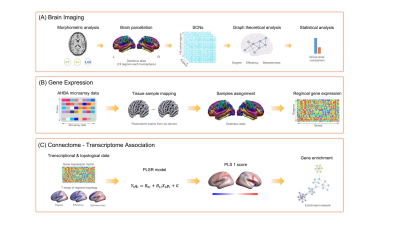 |
Brain connectomic and transcriptional signatures of suicidal
thoughts and behaviors in major depressive disorder
Kun Qin1,2,
Huiru Li3,
Rui Hu1,
Lisha Zhang2,
Cunqing Kong1,
Wen Chen1,
Qiyong Gong2,
and Zhiyun Jia2
1Department of Radiology, Taihe Hospital, Hubei University of Medicine, Shiyan, China, 2West China Hospital of Sichuan University, Chengdu, China, 3The First Affiliated Hospital of Kunming Medical University, Kunming, China Keywords: Psychiatric Disorders, Brain Motivation: Suicide-related connectomic signatures in depression and underlying transcriptional patterns have been poorly understood, most previous findings were limited by small-sample and single-site design. Goal(s): To identify robust brain structural network deficits associated with suicidal thoughts and behaviors (STB) in major depressive disorder (MDD) and to determine related transcriptional profiles. Approach: Based on mutlicenter MRI data of over 700 individuals, group-level connectomic comparisons and connectome-transcriptome association were analyzed. Results: Robust structural connectomic alterations associated with STB in MDD were distributed in the prefrontal, limbic and temporal areas. STB-related connectomic alterations were spatially correlated with genes enriched for cellular metabolism and synaptic signaling. Impact: These findings reveal a robust pattern of brain structural deficits at network level and demonstrate its linkage to gene expression patterns, which provides novel insights into the neurobiological underpinnings and potential markers for prediction and prevention of STB. |
14:45 |
1243.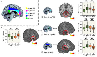 |
Effects of bright light therapy on cingulate cortex dynamic
functional connectivity and neurotransmitter activity in
subthreshold depression
Guixian Tang1,
Pan Chen1,
Guanmao Chen1,
Wei Cui2,
and Ying Wang1
1First Affiliated Hospital of Jinan University, Guangzhou, China, 2MR Research, GE Healthcare, Beijing, China, Guangzhou, China Keywords: Psychiatric Disorders, fMRI (resting state) Motivation: Bright light therapy (BLT) is one of the effective interventions for subthreshold depression, but its neural mechanism is still unclear. Goal(s): The goal of this double-blind, randomized, placebo-controlled clinical trial was to assess the correlation between BLT and the dynamic functional connectivity (dFC) changes in the cingulate cortex along with distribution of specific neurotransmitters in subthreshold depression. Approach: A double-blind randomized controlled trial Results: BLT alleviates depressive symptoms and changes the cingulate cortex dFC variability in subthreshold depression. And pre-treatment dFC variability of the cingulate cortex could be used as a biomarker for improved BLT treatment in subthreshold depression. Impact: BLT alleviates depressive symptoms and changes the cingulate cortex dFC variability in subthreshold depression, which raises the possibility that pre-treatment dFC variability of the cingulate cortex could be used as a biomarker for improved BLT treatment in subthreshold depression. |
| 14:57 |
1244.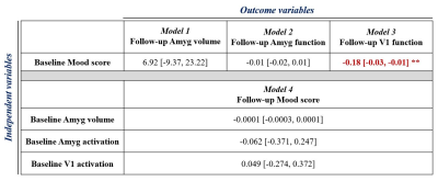 |
Current mood status is associated with future brain MRI
readouts, but not the other way around: insights from healthy
adults in the UK Biobank
Guocheng Jiang1,2,
Walter Swardfager2,3,
Hugo Cogo-Moreira4,
Sandra E Black2,5,
Benjamin I Goldstein3,6,
and Bradley J MacIntosh1,2
1Department of Medical Biophysics, University of Toronto, Toronto, ON, Canada, 2Hurvitz Brain Science Research Program, Sunnybrook Research Institute, Toronto, ON, Canada, 3Department of pharmacology and toxicology, University of Toronto, Toronto, ON, Canada, 4Department of Education, ICT and Learning, Østfold University College, Halden, Norway, 5Department of Medicine, University of Toronto, Toronto, ON, Canada, 6Centre for Youth Bipolar Disorder, The Centre for Addiction and Mental Health, Toronto, ON, Canada Keywords: Psychiatric Disorders, Psychiatric Disorders, Mood Motivation: The mood status of an individual can influence the brain anatomy and function. Goal(s): We studied whether the mood state is associated with brain structural and functional alteration at 2.25 years follow-up and vice versa. Approach: We focus on the regional brain volumes and task-induced function of the amygdala and primary visual cortex using T1w and task fMRI. Linear models tested for associations between mood and MRI readouts. Results: Baseline mood score is significantly associated with functional activation in the primary visual cortex. However, baseline volume and functional activation in the amygdala and primary visual cortex are not associated with future mood status. Impact: We showed that mood status is associated with future functional activation in the primary visual cortex. However, MRI estimates of anatomy and function in the amygdala and primary visual cortex demonstrated a lack of predictive power for future mood status. |
| 15:09 |
1245.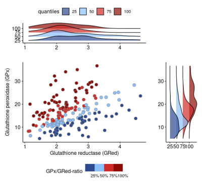 |
Associations between blood markers of redox regulation and brain
white matter microstructure display distinct signatures in
psychosis
Tommaso Pavan1,2,
Yasser Alemán-Gómez1,2,
Pascal Steullet1,
Zoe Schilliger1,3,
Daniella Dwir1,
Raoul Jenni1,3,
Martine Cleusix1,
Luis Alameda1,3,
Kim Q. Do1,3,
Philippe Conus1,
Paul Klauser1,3,
Patric Hagmann1,
and Ileana Jelescu1
1Lausanne University Hospital (CHUV), Lausanne, Switzerland, 2University of Lausanne, Lausanne, Switzerland, 3University of Lausanne (UNIL), Lausanne, Switzerland Keywords: Psychiatric Disorders, White Matter, biomarker, diffusion, DKI, kurtosis, DTI, DWI, psychosis Motivation: It is unclear whether Glutathione peroxidase (GPx) and reductase (GRed) activity, peripheral proxy of redox homeostasis, could be linked to white matter alterations in psychosis.
Goal(s): We seek to link white matter alterations to
processes connected to neuroinflammation, namely raising
from dysregulation of the glutathione redox homeostasis.
Approach: We applied diffusion kurtosis imaging and
White Matter Tract Integrity – Watson to patients data and
analyzed the maps via Tract-Based Spatial Statistics(TBSS)
and regression contrast analysis.
Results: We found selective and widespread
association of kurtosis metrics with glutathione redox
enzymes that significantly dissociate between patients and
controls.
Impact: Our findings reveal that Redox GSH
dysregulation in patients may impact WM microstructure,
indicating therapeutic possibilities and reinforcing the
connection between microstructure and neuroinflammation.
Furthermore, the neat selectivity of the kurtosis metrics
highlights potential for widespread clinical research
applications. |
| 15:21 |
1246.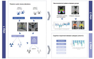 |
Two distinct thalamic subtypes associated with cognitive
impairment in first-episode schizophrenia
Wei Yu1,2,
Bo Tao1,2,
Qiannan Zhao2,3,
Yuan Xiao1,2,
Fei Zhu1,2,
Ziyang Gao1,2,
and Su Lui1,2
1Department of Radiology, West China Hospital of Sichuan University, Chengdu, China, 2Research Unit of Psychoradiology, Chinese Academy of Medical Sciences, Chengdu, China, 3Huaxi MR Research Center (HMRRC), West China Hospital of Sichuan University, Chengdu, China Keywords: Psychiatric Disorders, Brain, thalamic nuclei Motivation: Schizophrenia is characterized by high heterogeneity, with the core symptom of cognitive impairment. However, it is unclear that whether the patterns of thalamic nuclei volume alterations is associated with cognitive function in schizophrenia. Goal(s): To investigate the pattern and heterogeneity of thalamic nuclei alterations and cognition in schizophrenia. Approach: The cluster analysis (K-means++) was used to identify the subtype of schizophrenia. And machine learning model was developed to validate the reproducibility of subtypes. Results: We uncovered two markedly distinct neuroanatomical subtypes of schizophrenia. One of the subtypes characterized as widespread decrease in thalamic nuclei and severe cognition deficit. Impact: These findings could help to identify biological targets related to the treatment of schizophrenia, and improve the functional outcomes in patients with schizophrenia. |
 Back to Meeting Home
 Back to the Program-at-a-Glance
The International Society for Magnetic Resonance in Medicine is accredited by the Accreditation Council for Continuing Medical Education to provide continuing medical education for physicians.
 View Presentation Video